
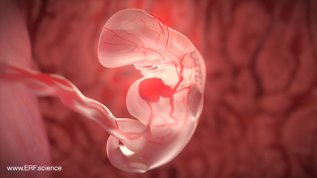
None of the medical imagery used by the Education Resource Fund (ERF) to depict embryonic and fetal development was created using generative artificial intelligence (AI) or any other animation process.
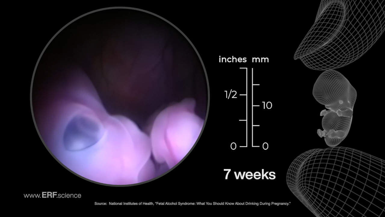
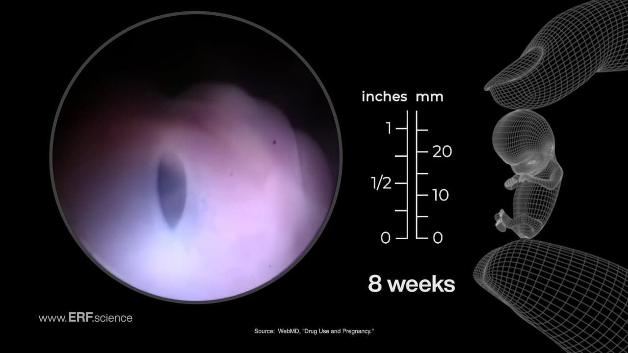
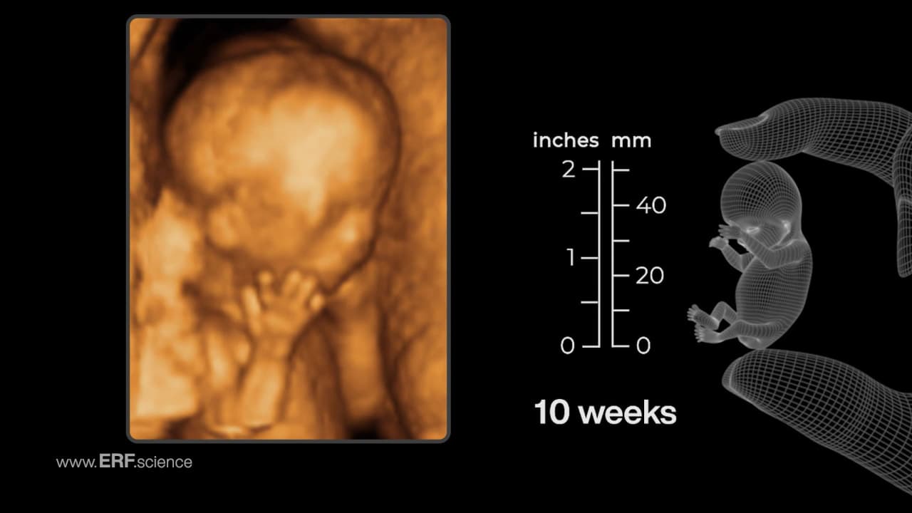
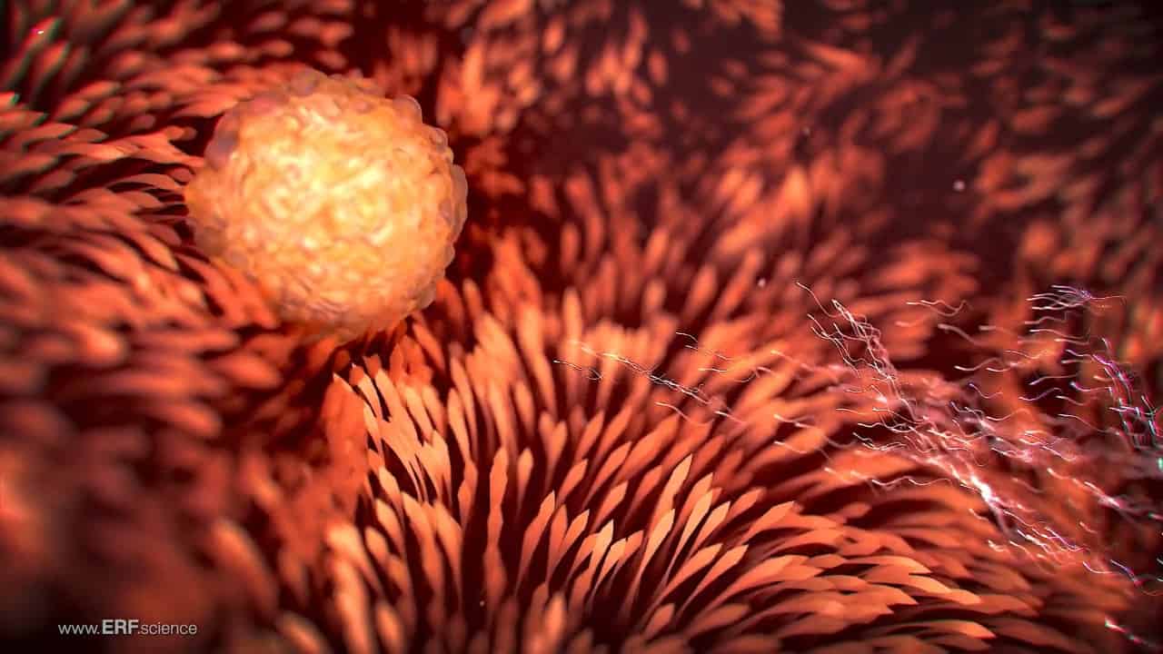
Imagery featured on this site was derived primarily through endoscopic scans, with secondary content produced by sonographic scans, high-magnification light microscopy, electron microscopy, and magnetic resonance imaging, all depicting actual human embryos and fetuses.
The authenticity of all content on the ERF website and in the ERF app can be verified by reference to the original tapes recorded by the physicians conducting the featured diagnostic and therapeutic scans.
Astonishing embryonic and fetal endoscopic video shorts are now available in formats which enable posting on all major social media platforms.
Contact the Education Resource Fund (ERF) for licensing information, including pricing and terms of use.
LANDSCAPE CONFIGURATION (HORIZONTAL)
PORTRAIT CONFIGURATION (VERTICAL)
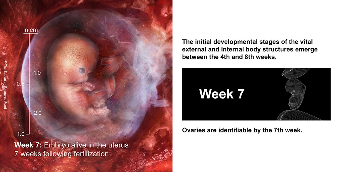
3D Organ Model Video
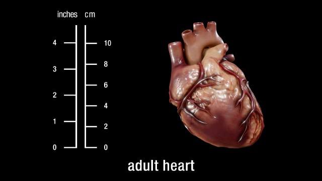
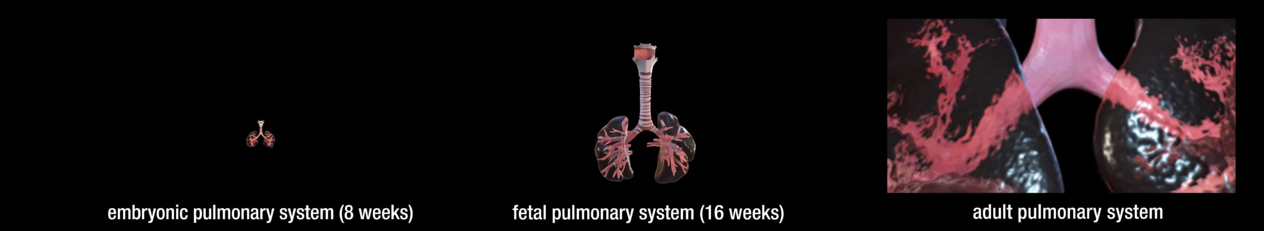
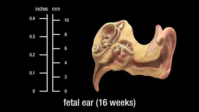
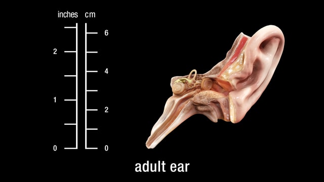
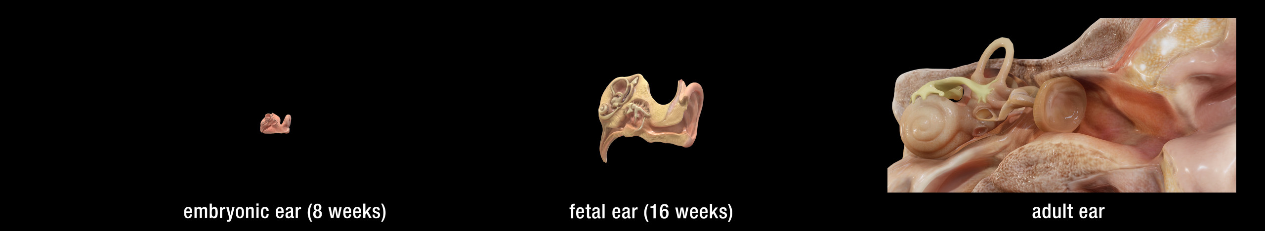
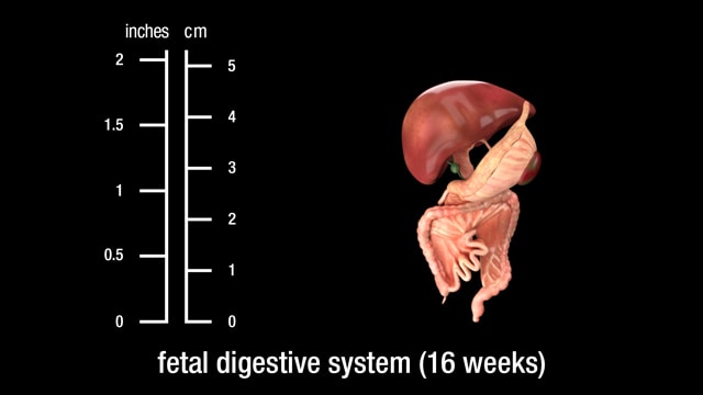
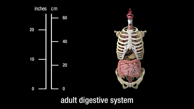
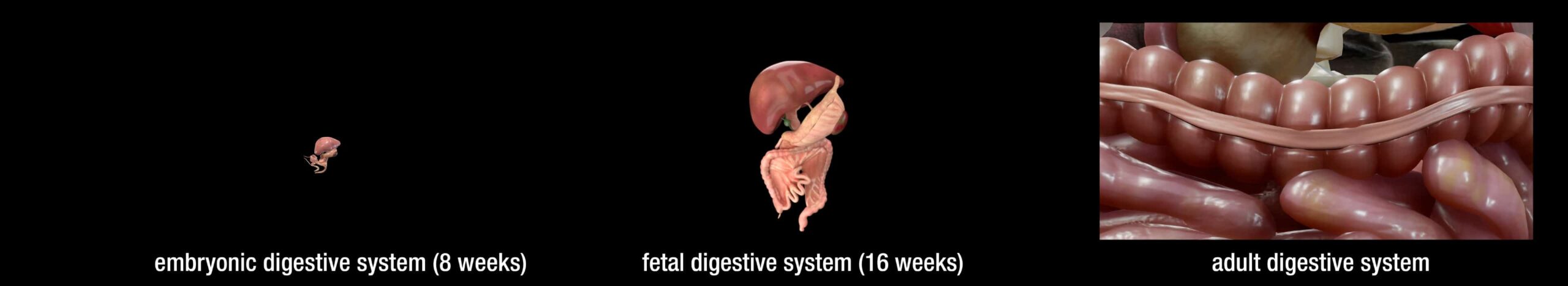
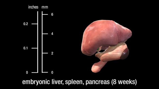
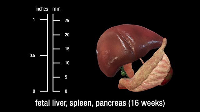
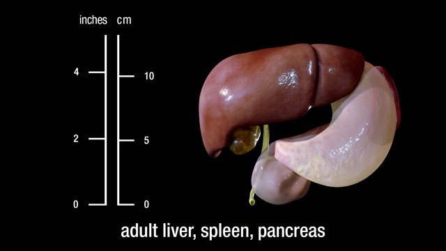
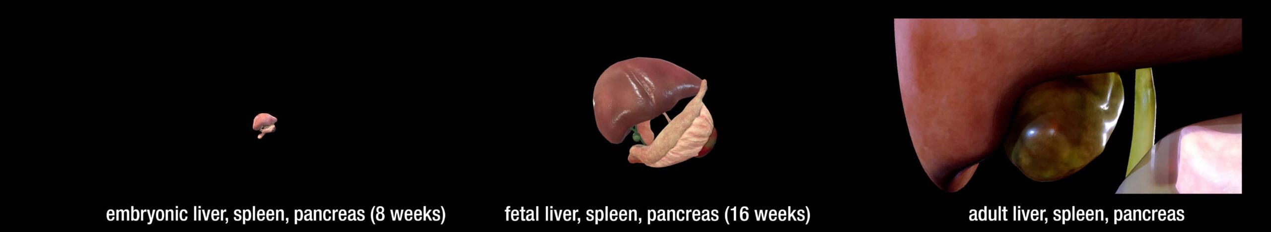
Comparative Embryonic and Fetal Anatomy
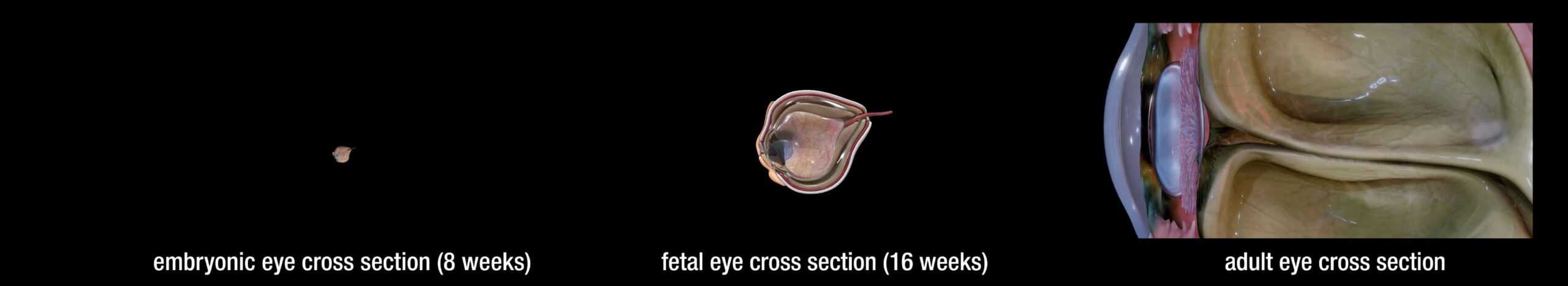
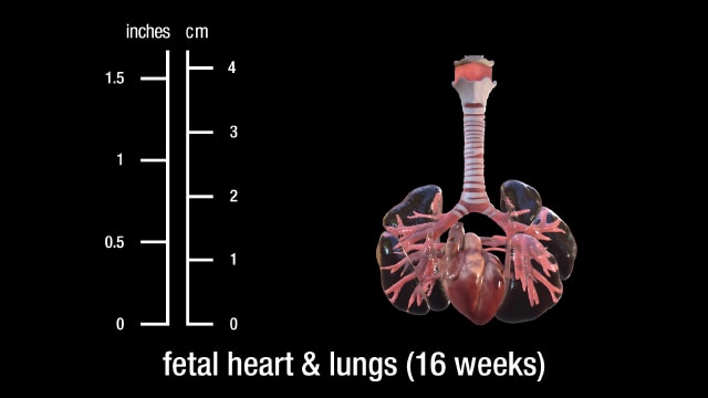
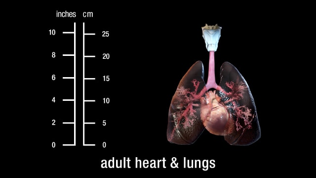
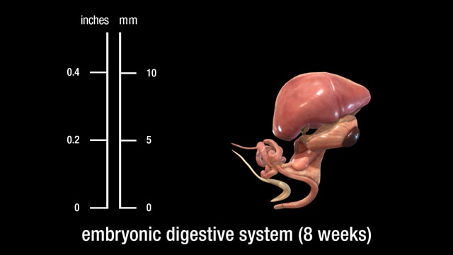
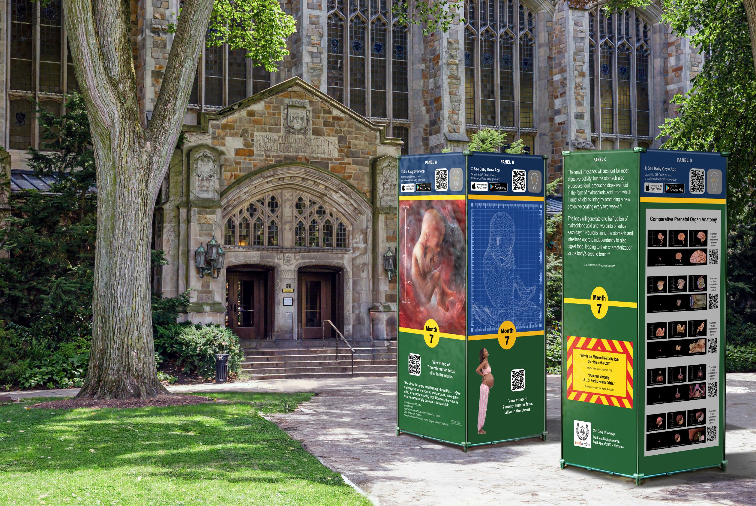
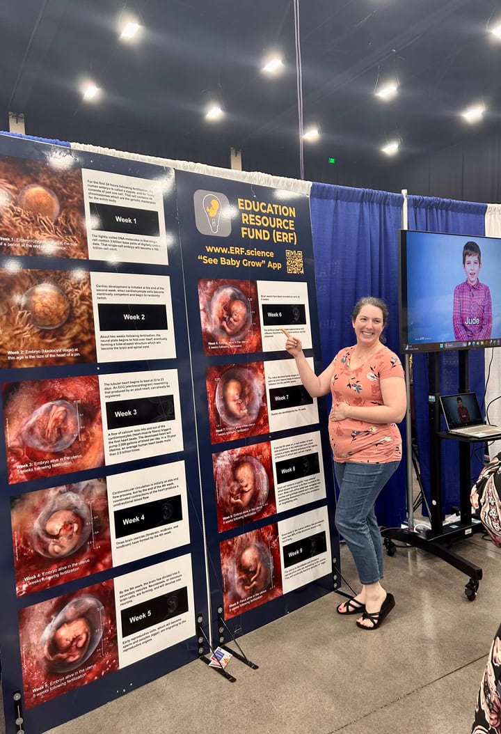
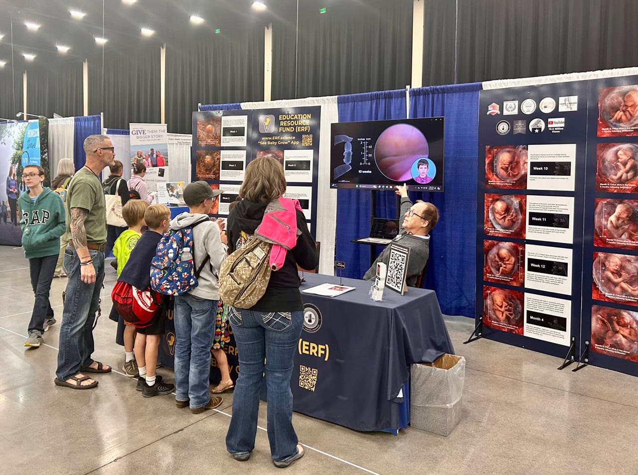
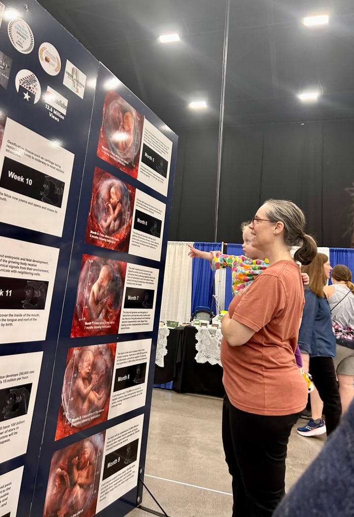
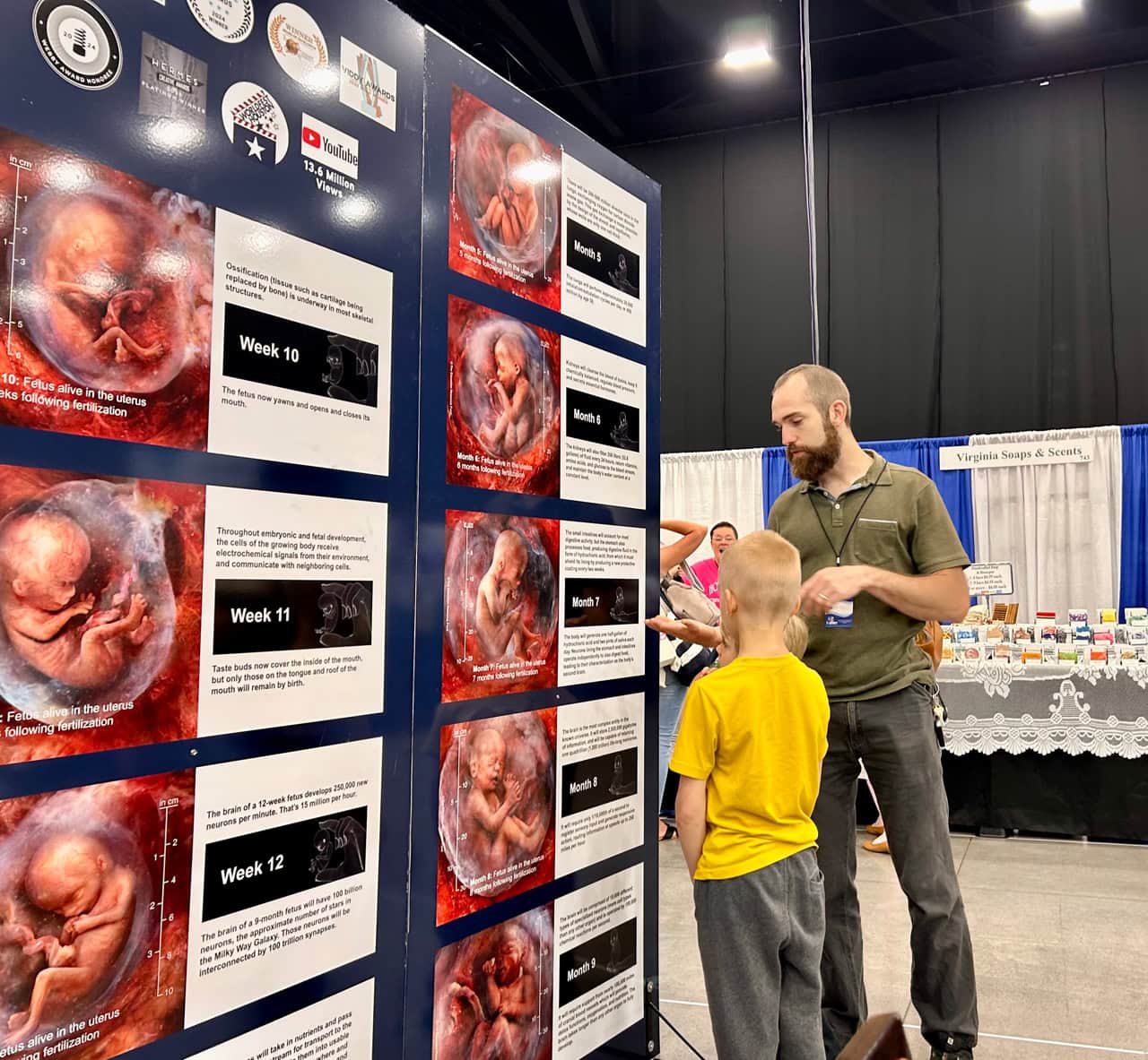
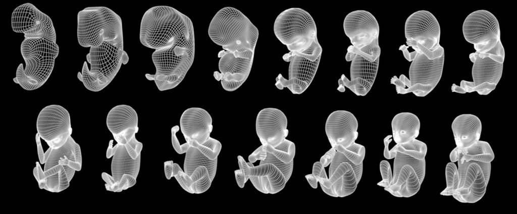
See 12 of your baby's vital organs depicted in embryonic, fetal, and adult age stages and illustrated as 3D models which you can rotate about their vertical, horizontal, etc. axes
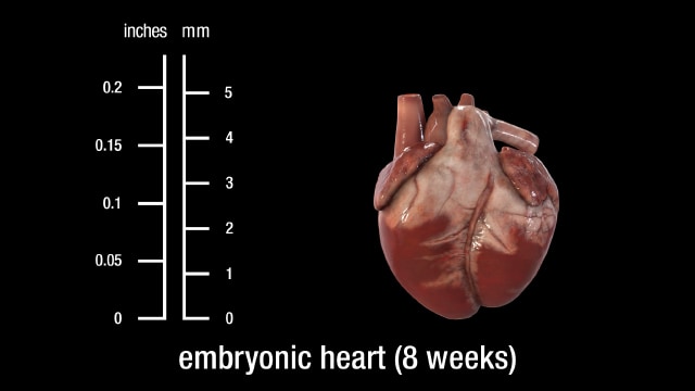
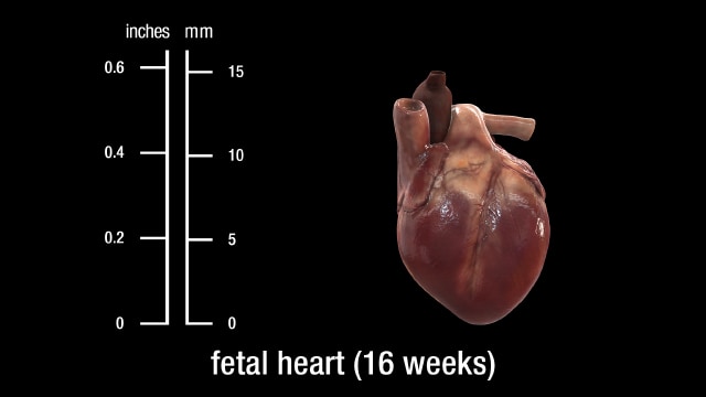
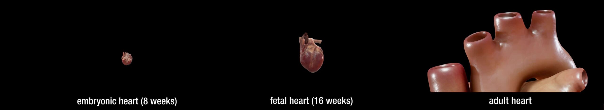

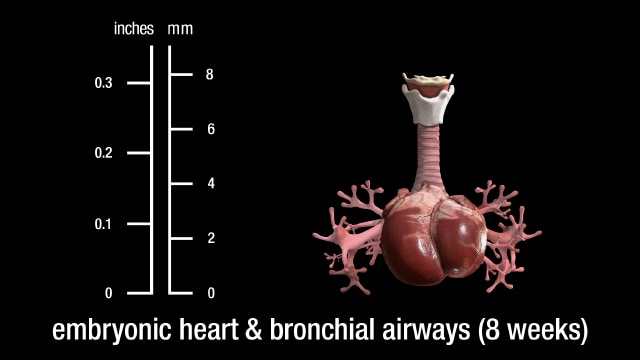
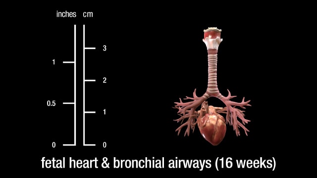
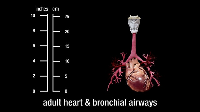
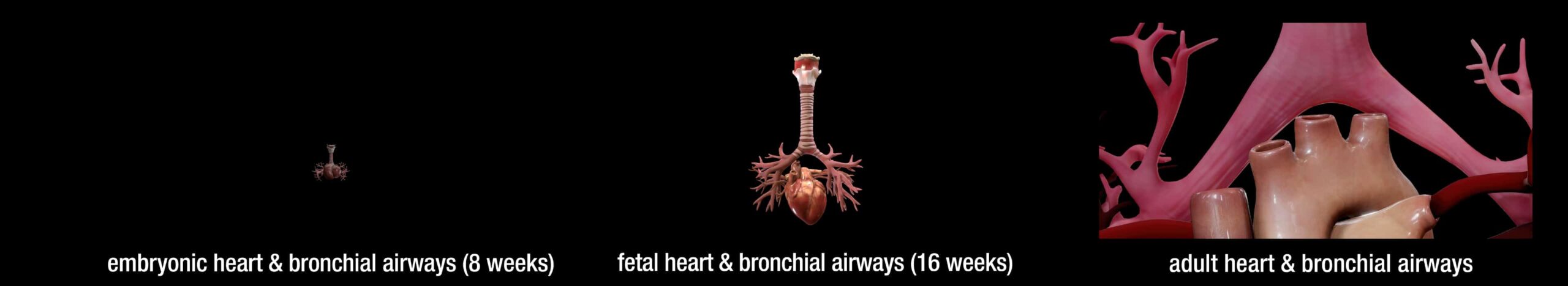
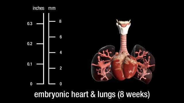
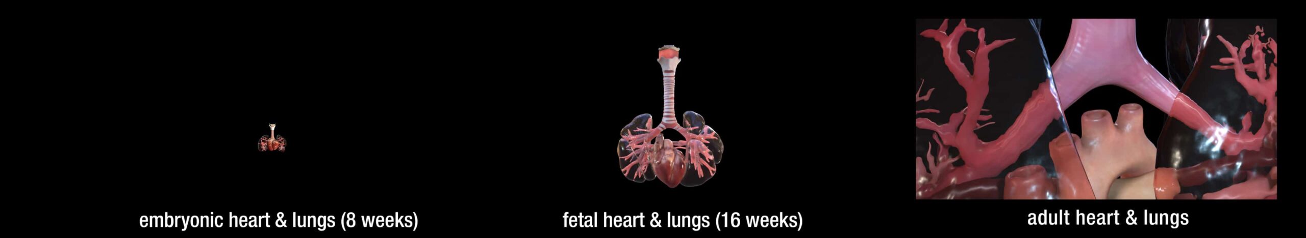
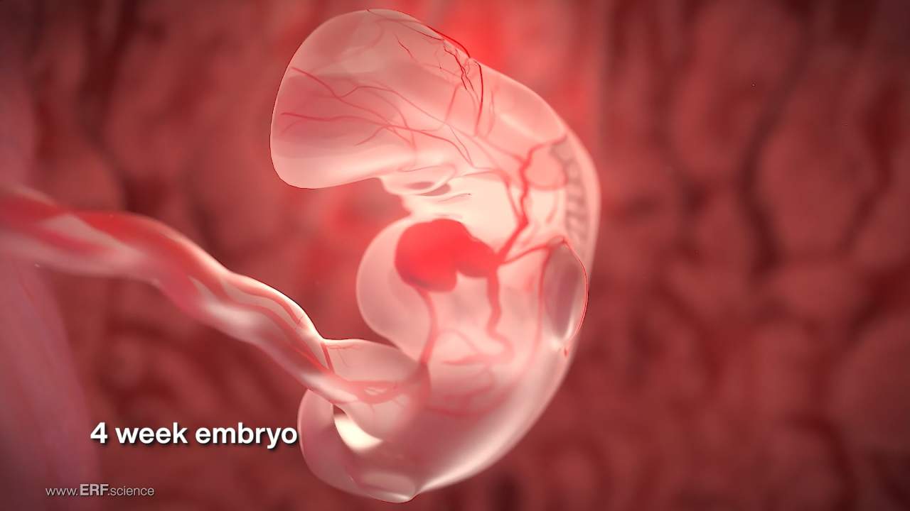
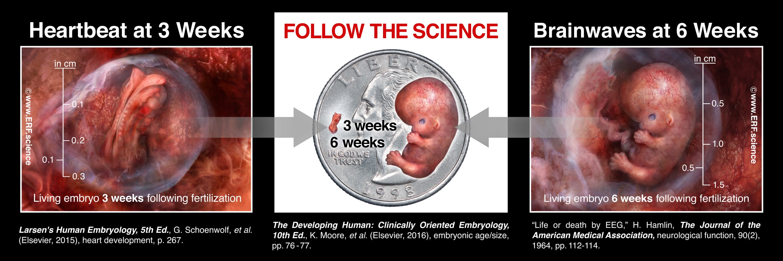
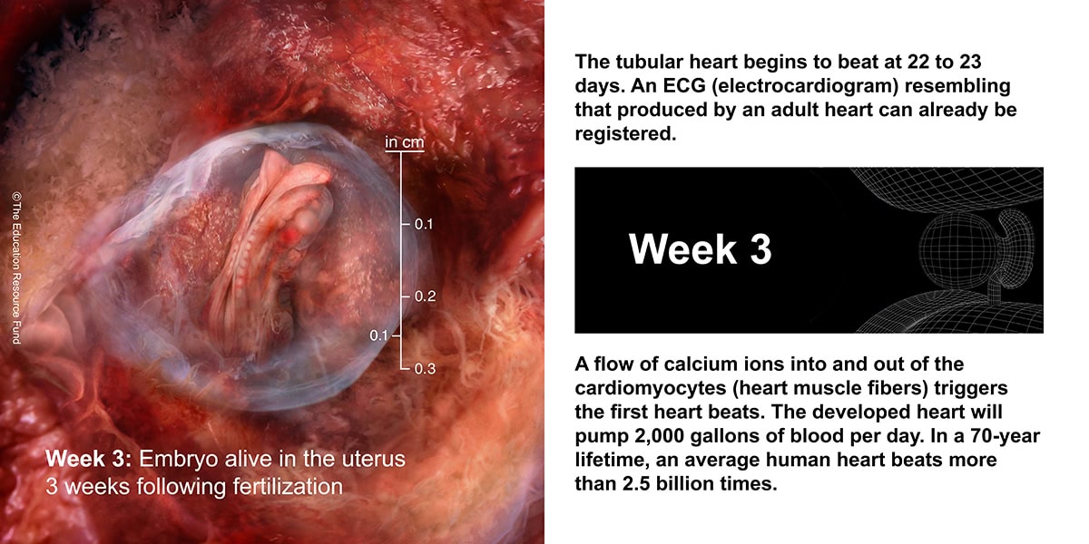
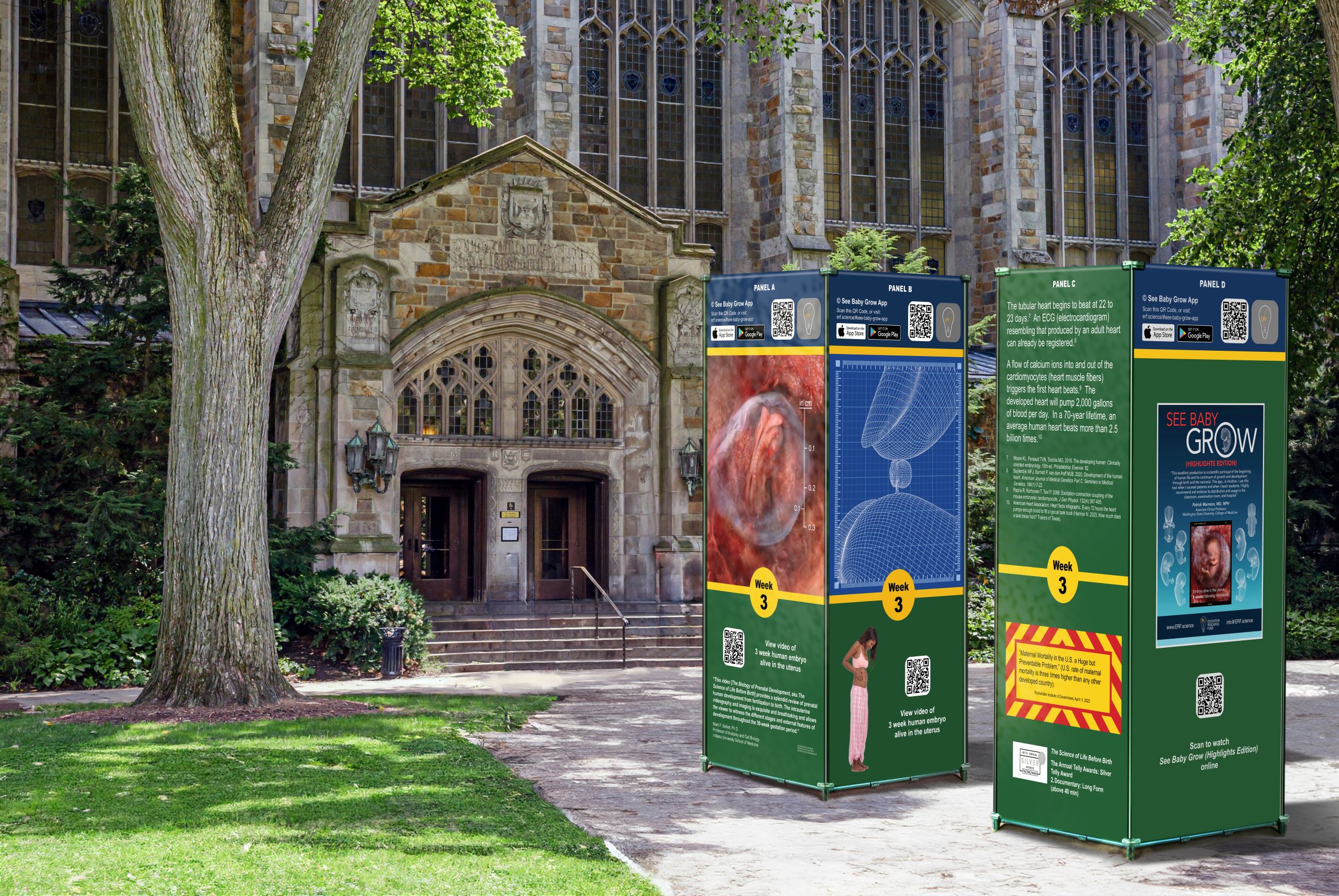
The heart is the first functional organ in vertebrate embryos.
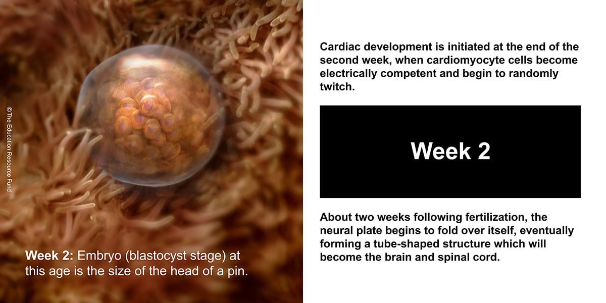
Heart development (also known as cardiogenesis) refers to the prenatal development of the heart. This begins with the formation of two endocardial tubes which merge to form the tubular heart, also called the primitive heart tube. The heart is the first functional organ in vertebrate embryos.
The heart is a muscular organ in most animals, which pumps blood through the blood vessels of the circulatory system. Blood provides the body with oxygen and nutrients, as well as assisting in the removal of metabolic wastes. In humans, the heart is located between the lungs, in the middle compartment of the chest.
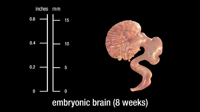
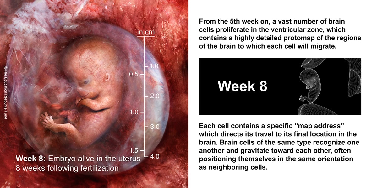
During weeks 8 to 10, the cerebrum begins its development in earnest. Neurons proliferate and begin their migration throughout the brain. The anterior commissure, which is the first interhemispheric connection (a small one), also develops. Reflexes appear for the first time during this period.
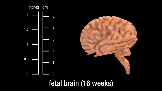
In the second trimester, the fetal brain begins to direct the compression of the chest muscles and movement of the diaphragm. These are like practice breaths and are controlled by the brainstem. Sucking and swallowing begin around week 16, and by week 21 the fetus can swallow amniotic fluid.
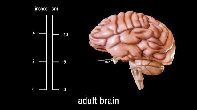
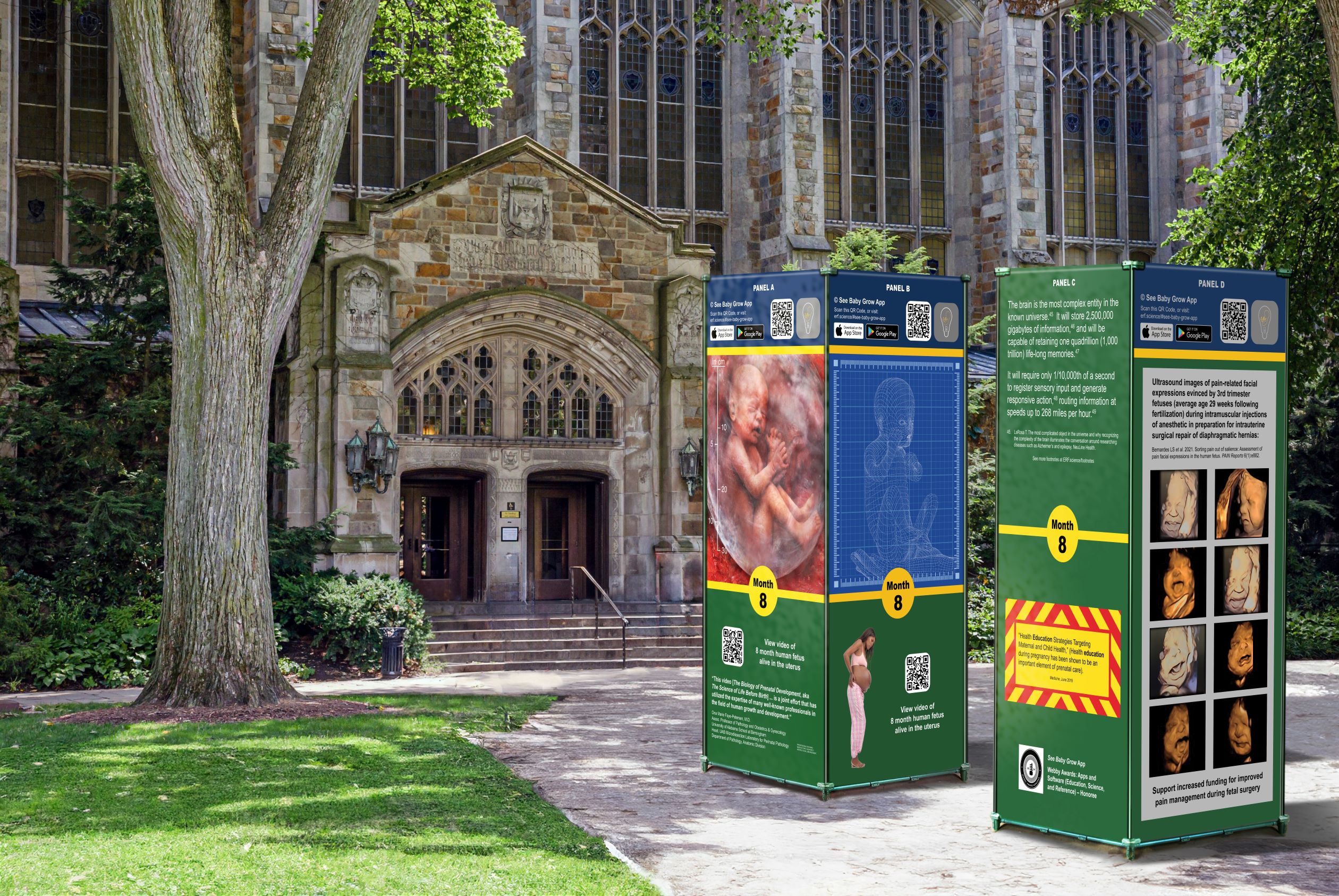
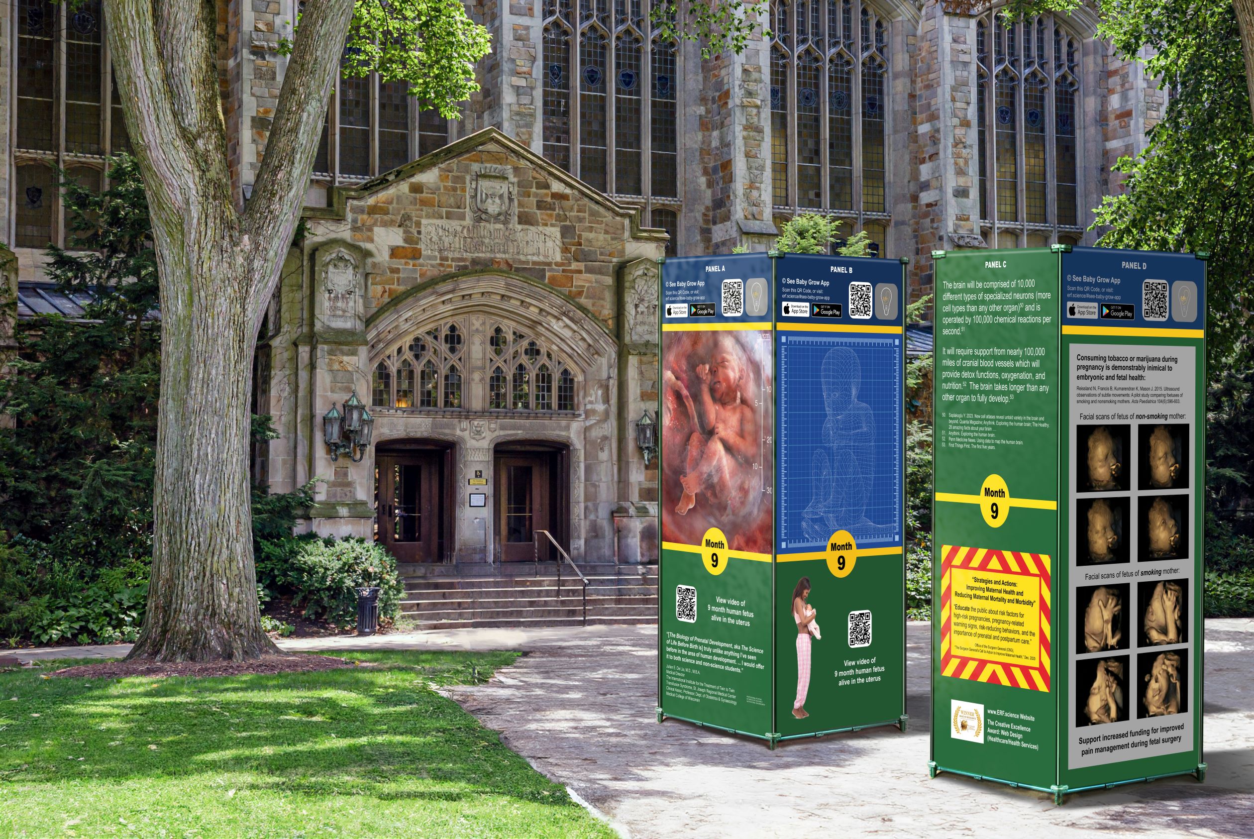
The brainstem provides the main motor and sensory nerve supply to the face and neck via the cranial nerves. Of the thirteen pairs of cranial nerves, ten pairs (or twelve, if the diencephalon is included in the brainstem) come from the brainstem. The brainstem is an extremely important part of the brain as the nerve connections of the motor and sensory systems from the main part of the brain to the rest of the body pass through the brainstem. This includes the corticospinal tract (motor), the dorsal column-medial lemniscus pathway (fine touch, vibration sensation, and proprioception), and the spinothalamic tract (pain, temperature, itch, and crude touch). The human brain is the central organ of the human nervous system, and with the spinal cord makes up the central nervous system.
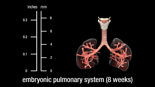
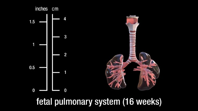
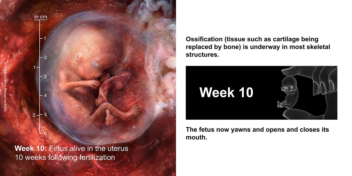
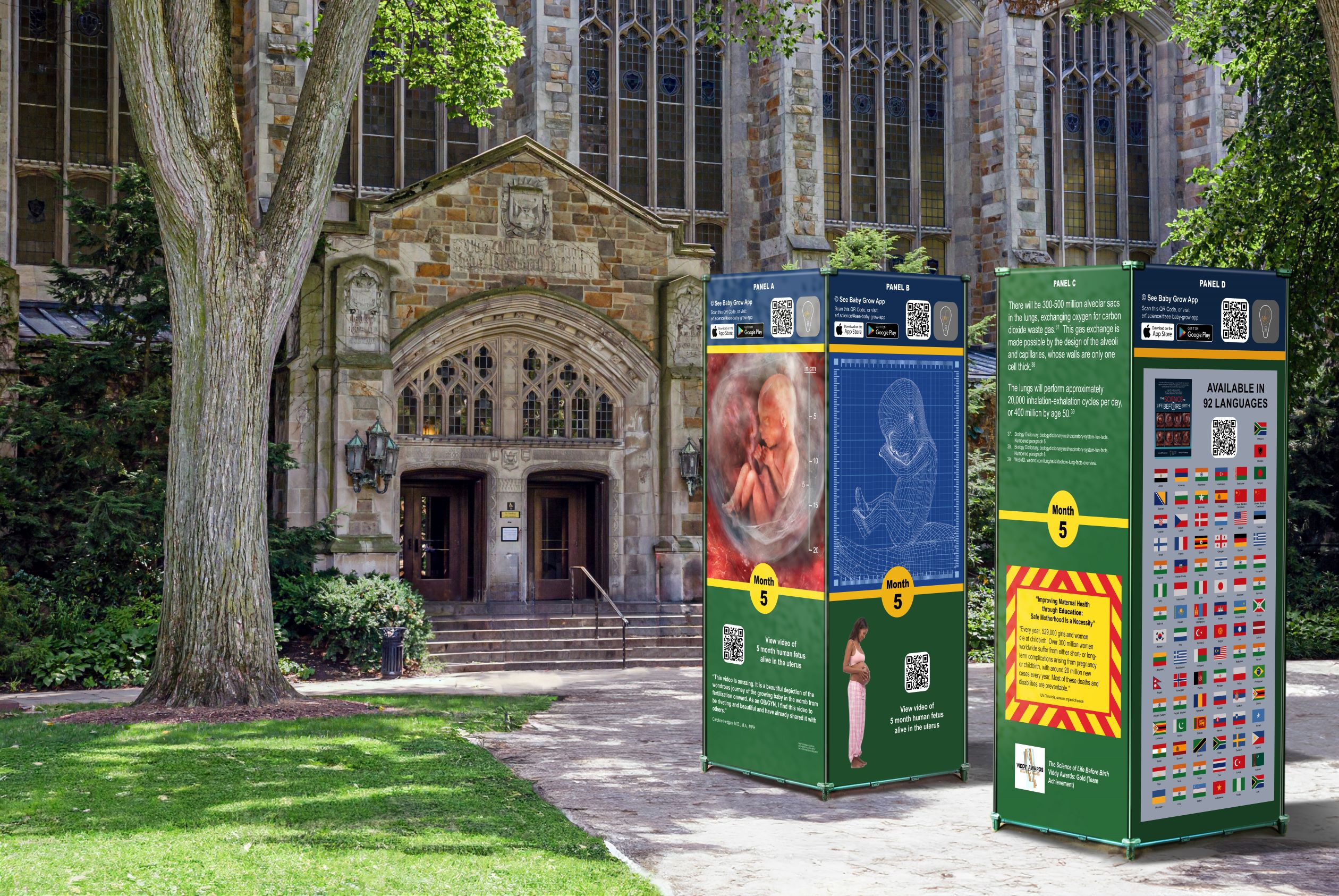
Stages of normal human lung development: During the first stage (0-7 weeks, embryonic stage), the lung arises as a ventral diverticulum of the primitive foregut endoderm with the lobar and segmental bronchi appearing at the fifth week and arteries and veins developing as avascular buds. There then follows the pseudoglandular stage (8-17 weeks) when branching of the airways and vessels takes place.
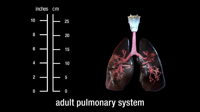
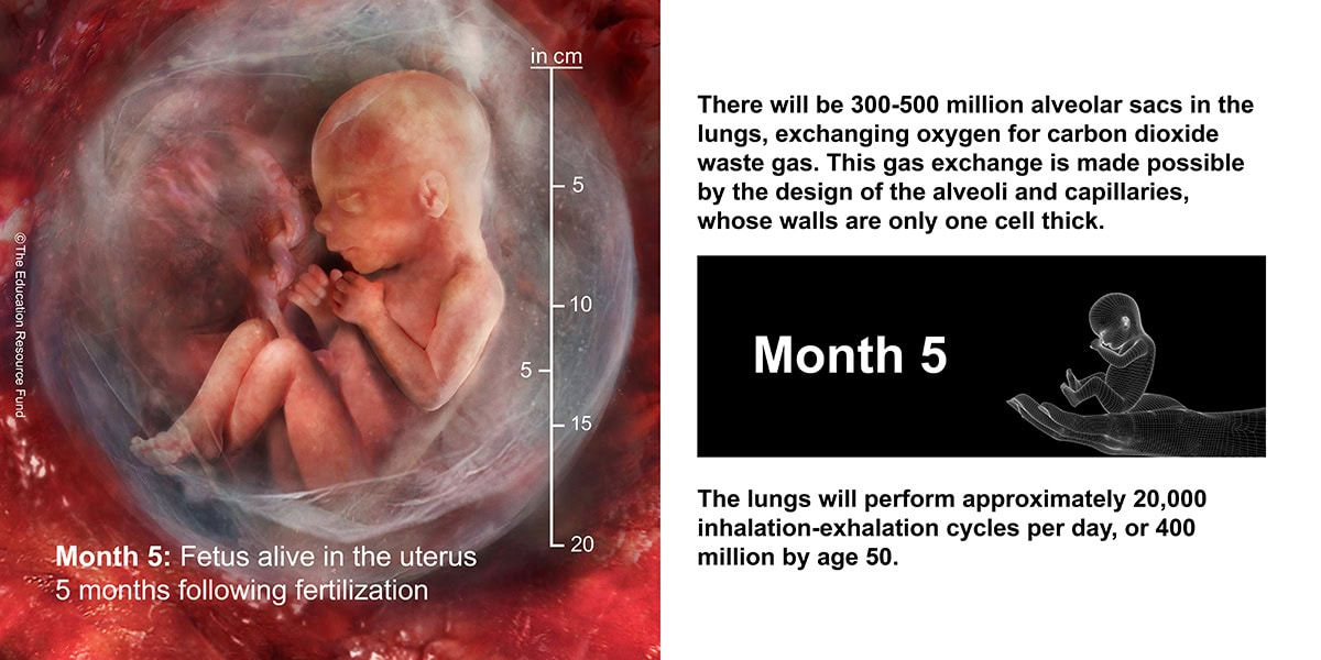
The respiratory system has conducting and gas exchange components, with the bifurcating airways and accompanying pulmonary arteries conducting air and blood to peripheral capillary-lined airspaces for gas exchange.
The pulmonary system includes the lungs, the bronchial tree, and the pulmonary vessels. The typical respiratory rate for a healthy adult at rest is 12–18 breaths per minute. The respiratory center sets the quiet respiratory rhythm at around two seconds for an inhalation and three seconds exhalation. This gives the lower of the average rate at 12 breaths per minute.
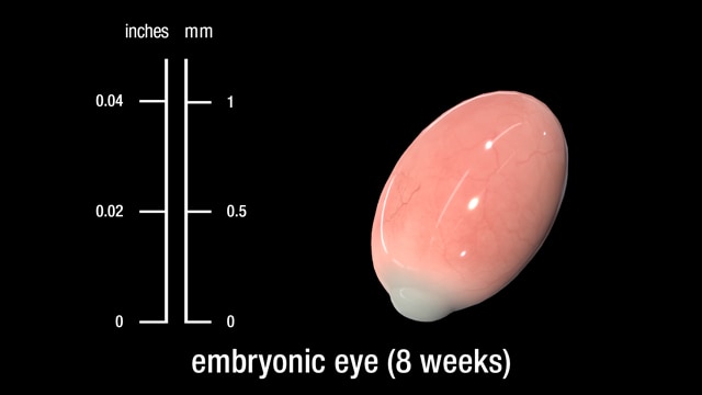
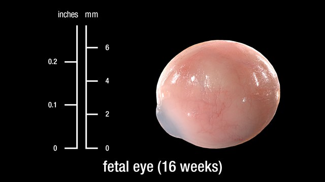
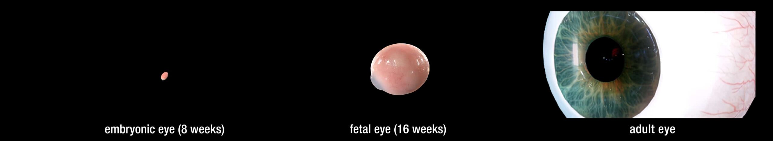
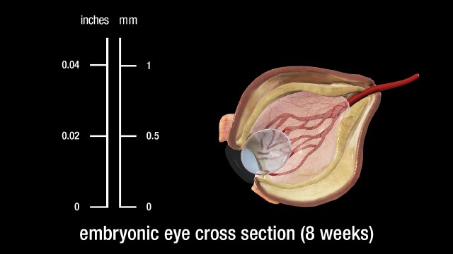
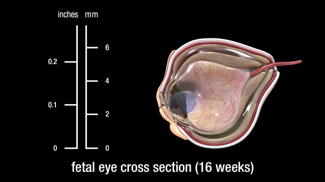
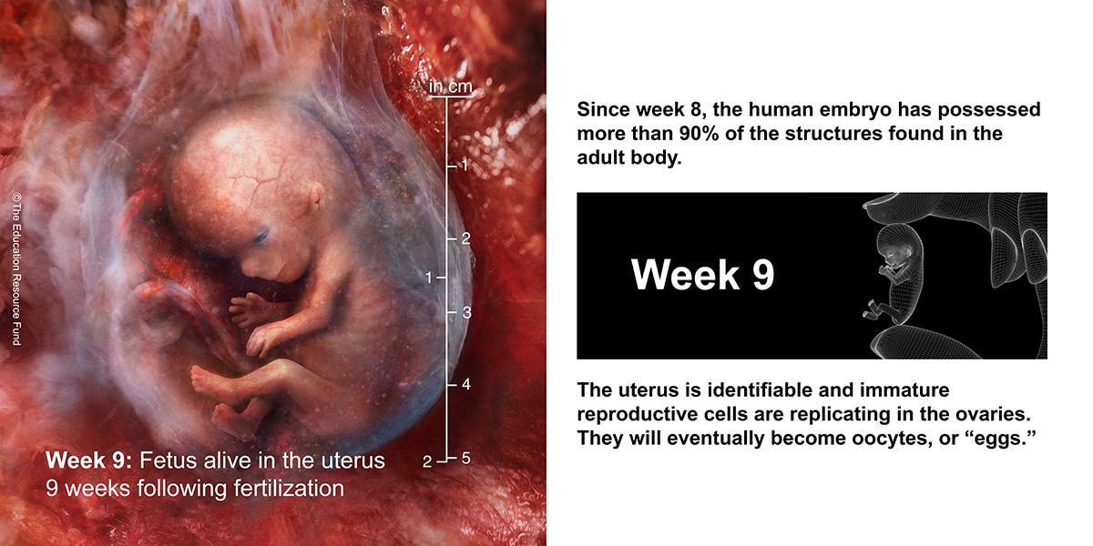
The eyes have moved forward on the face. At 8 weeks, the eyelids start to form and fuse together to protect the other developing eye structures.
Although eye development can be considered to commence around day 22, when the optic sulci (optic primordium) appear as shallow grooves or pits in the inner aspect of the neural plate or neural folds and the embryo is around 2 mm in length with eight somites, the group of cells that constitute the eye primordium or eye field have already begun to express a set of eye field transcription factors (EFTFs).
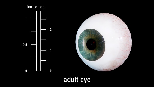
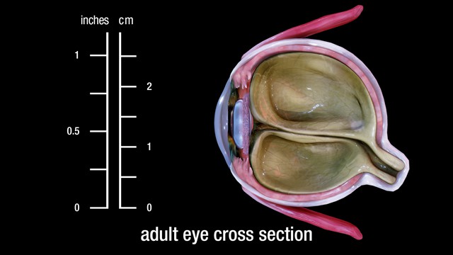
The human eye is a sensory organ, part of the sensory nervous system, that reacts to visible light and allows us to use visual information for various purposes including seeing things, keeping our balance, and maintaining circadian rhythm.
Cells differentiate into specific types according to their projected role in the visual process, such as rods and cones, with axons from the retinal ganglion cells forming the optic nerve. It takes six months for the retinal layers to grow from the neural ectoderm. The macula needs four or five months just to begin, and it will mature six months after birth.
Although eye development can be considered to commence around day 22, when the optic sulci (optic primordium) appear as shallow grooves or pits in the inner aspect of the neural plate or neural folds and the embryo is around 2 mm in length with eight somites, the group of cells that constitute the eye primordium or eye field have already begun to express a set of eye field transcription factors (EFTFs).
Eyes are organs of the visual system. They provide living organisms with vision, the ability to receive and process visual detail, as well as enabling several photo response functions that are independent of vision. Eyes detect light and convert it into electrochemical impulses in neurons. In higher organisms, the eye is a complex optical system which collects light from the surrounding environment, regulates its intensity through a diaphragm, focuses it through an adjustable assembly of lenses to form an image, converts this image into a set of electrical signals, and transmits these signals to the brain through complex neural pathways that connect the eye via the optic nerve to the visual cortex and other areas of the brain.
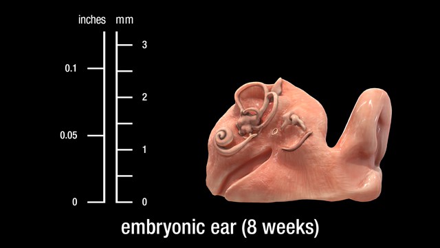
The ear is a composite structure with multiple embryonic origins , the external and middle ears arise from the first and second pharyngeal arches and the intervening pharyngeal cleft , membrane and pouch.
The ear is a composite structure with multiple embryonic origins. The external and middle ears arise from the first and second pharyngeal arches and the intervening pharyngeal cleft, membrane, and pouch.
The ear is the organ that enables hearing and, in mammals, body balance using the vestibular system. In mammals, the ear is usually described as having three parts—the outer ear, the middle ear and the inner ear. The outer ear consists of the pinna and the ear canal. The middle ear includes the tympanic cavity and the three ossicles. The inner ear sits in the bony labyrinth, and contains structures which are key to several senses: the semicircular canals, which enable balance and eye tracking when moving; the utricle and saccule, which enable balance when stationary; and the cochlea, which enables hearing. The ears of vertebrates are placed somewhat symmetrically on either side of the head, an arrangement that aids sound localization.
Heart development (also known as cardiogenesis) refers to the prenatal development of the heart. This begins with the formation of two endocardial tubes which merge to form the tubular heart, also called the primitive heart tube. The heart is the first functional organ in vertebrate embryos. The respiratory system has conducting and gas exchange components, with the bifurcating airways and accompanying pulmonary arteries conducting air and blood to peripheral capillary-lined airspaces for gas exchange.
Heart development (also known as cardiogenesis) refers to the prenatal development of the heart. This begins with the formation of two endocardial tubes which merge to form the tubular heart, also called the primitive heart tube. The heart is the first functional organ in vertebrate embryos. The respiratory system has conducting and gas exchange components, with the bifurcating airways and accompanying pulmonary arteries conducting air and blood to peripheral capillary-lined airspaces for gas exchange.
A bronchus is a passage or airway in the lower respiratory tract that conducts air into the lungs. The first or primary bronchi to branch from the trachea at the carina are the right main bronchus and the left main bronchus. These are the widest bronchi, and enter the right lung and the left lung at each hilum. The main bronchi branch into narrower secondary bronchi or lobar bronchi, and these branch into narrower tertiary bronchi or segmental bronchi. Further divisions of the segmental bronchi are known as 4th order, 5th order, and 6th order segmental bronchi, or grouped together as subsegmental bronchi. The bronchi, when too narrow to be supported by cartilage, are known as bronchioles. No gas exchange takes place in the bronchi.
Stages of normal human lung development: During the first stage (0-7 weeks, embryonic stage), the lung arises as a ventral diverticulum of the primitive foregut endoderm with the lobar and segmental bronchi appearing at the fifth week and arteries and veins developing as avascular buds. There then follows the pseudoglandular stage (8-17 weeks) when branching of the airways and vessels takes place.
Heart development (also known as cardiogenesis) refers to the prenatal development of the heart. This begins with the formation of two endocardial tubes which merge to form the tubular heart, also called the primitive heart tube. The heart is the first functional organ in vertebrate embryos. The respiratory system has conducting and gas exchange components, with the bifurcating airways and accompanying pulmonary arteries conducting air and blood to peripheral capillary-lined airspaces for gas exchange.
The normal resting adult heart rate is 60–100 beats per minute. The typical respiratory rate for a healthy adult at rest is 12–18 breaths per minute. The respiratory center sets the quiet respiratory rhythm at around two seconds for an inhalation and three seconds exhalation. This gives the lower of the average rate at 12 breaths per minute.
Both the epithelium and the parenchyma of glands associated with the digestive tract (e.g., liver and pancreas) are derived from endoderm. The muscular walls of the digestive tract (lamina propria, muscularis mucosae, submucosa, muscularis externa, adventitia and/or serosa) are derived from splanchnic mesoderm.
As a result of cephalocaudal and lateral folding of the embryo, a portion of the endoderm derived from gastrulation is incorporated into the embryo to form the primitive gut.
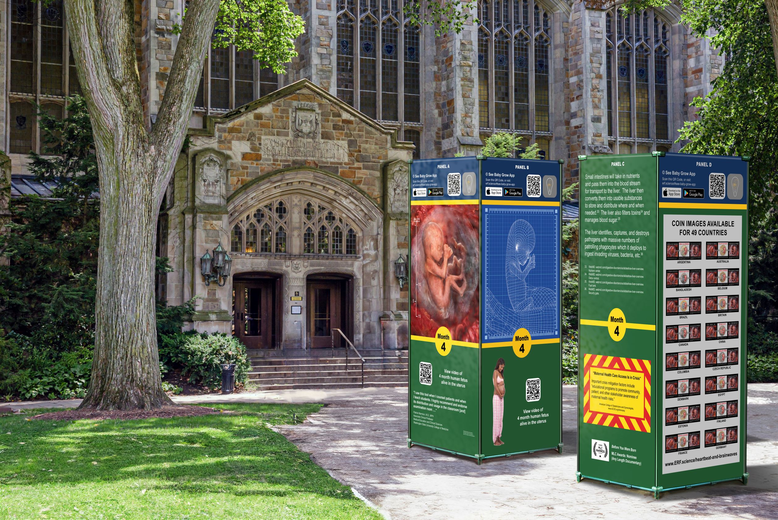
The digestive system plays a fundamental role ensuring liquids we ingest are broken down into useful nutrients and chemicals. The digestive tract consists of the mouth, or oral cavity, with its teeth, for grinding the food, and its tongue, which serves to knead food and mix it with saliva. Then, there is the throat, or pharynx; the esophagus; the stomach; the small intestine, consisting of the duodenum, the jejunum, and the ileum; and the large intestine, consisting of the cecum, a closed-end sac connecting with the ileum, the ascending colon, the transverse colon, the descending colon, and the sigmoid colon, which terminates in the rectum. Glands contributing digestive juices include the salivary glands, the gastric glands in the stomach lining, the pancreas, and the liver and its adjuncts – the gallbladder and bile ducts.
Both the epithelium and the parenchyma of glands associated with the digestive tract (e.g., liver and pancreas) are derived from endoderm. The muscular walls of the digestive tract (lamina propria, muscularis mucosae, submucosa, muscularis externa, adventitia and/or serosa) are derived from splanchnic mesoderm.
The liver, gallbladder, and biliary duct system arise as a ventral outgrowth from the caudal end of the foregut early in the fourth week of development. This outgrowth, known as the hepatic diverticulum, extends into the septum transversum, which is the future diaphragm. The hepatic diverticulum grows rapidly and divides into cranial and caudal portions. The larger cranial portion forms the primordium of the liver parenchyma. Proliferating endodermal cells develop into cords of hepatocytes and into the epithelial lining of the biliary system.
The pancreas and bile duct (biliary) systems together form an important part of the digestive system. The pancreas and liver produce juices (pancreatic juice and bile) which help in the process of digestion.
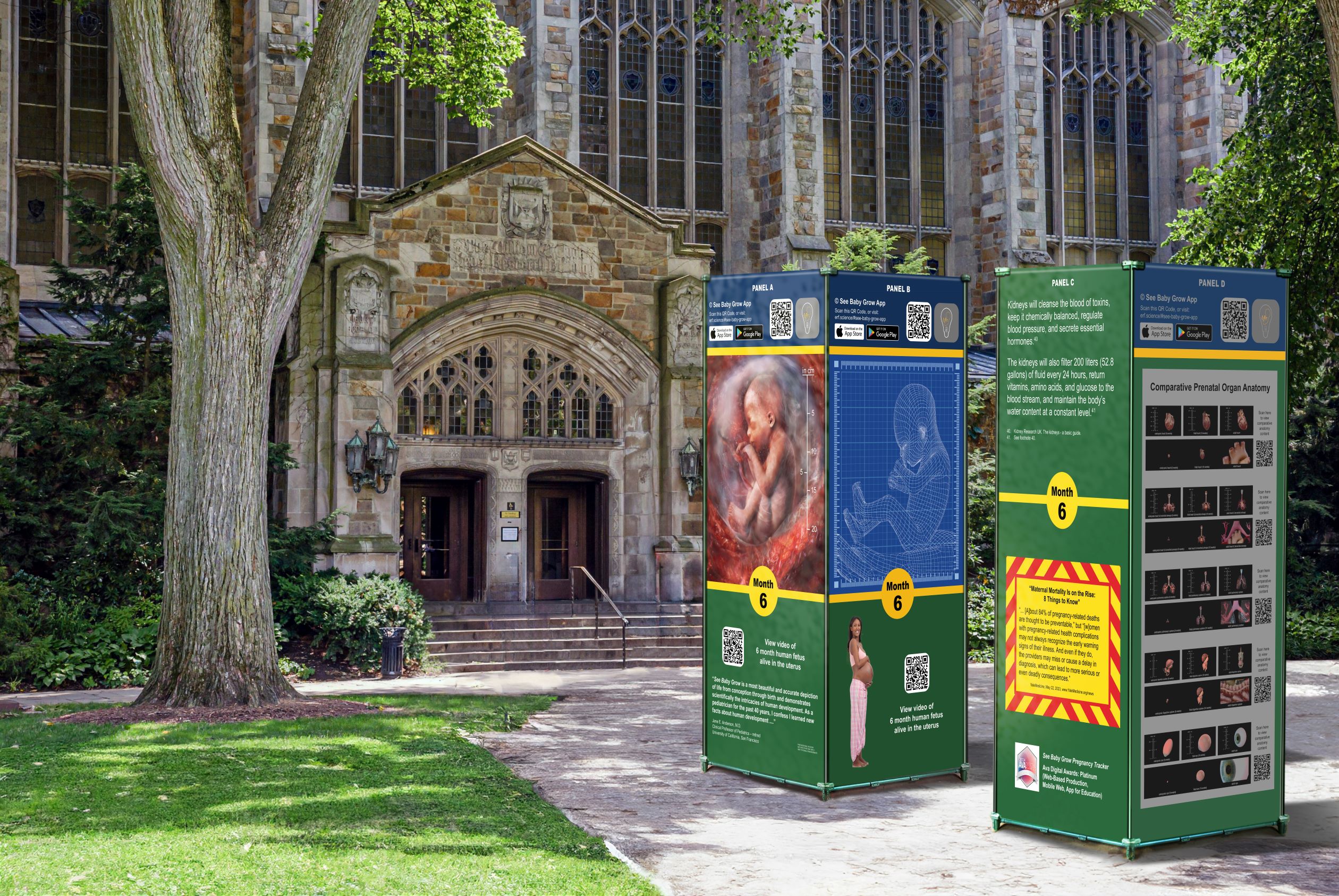
PROFESSIONAL ENDORSEMENTS OF THE EDUCATION RESOURCE FUND PRENATAL PROJECT
Professional reviews neither state nor imply institutional endorsement.
Principal Technical Advisor:
Mark T. Cullen, M.D.
Technical Review:
Enid Gilbert-Barness, M.D.
David H. Bernanke, Ph.D.
Mark J. Holterman, M.D., Ph.D.
David L. Bolender, Ph.D.
Paul A. Krieg, Ph.D.
Professor Stuart Campbell, D.Sc.
Maria Michejda, M.D,
Bruce M. Carlson, M.D., Ph.D.
Maurice J. Pescitelli Jr., Ph.D.
Charles L. Saxe, Ill, Ph.D.
Charles H. Ellis Jr., Ph.D.
Mark F. Seifert, Ph.D.
Ona Marie Faye-Petersen, M.D.
Allan R. Sinning, Ph.D.
David W. Fontaine, M.D.
Bradley R. Smith, Ph.D.
Ravmond F. Gasser, Ph.D.
Sam R. Voora, M.D.
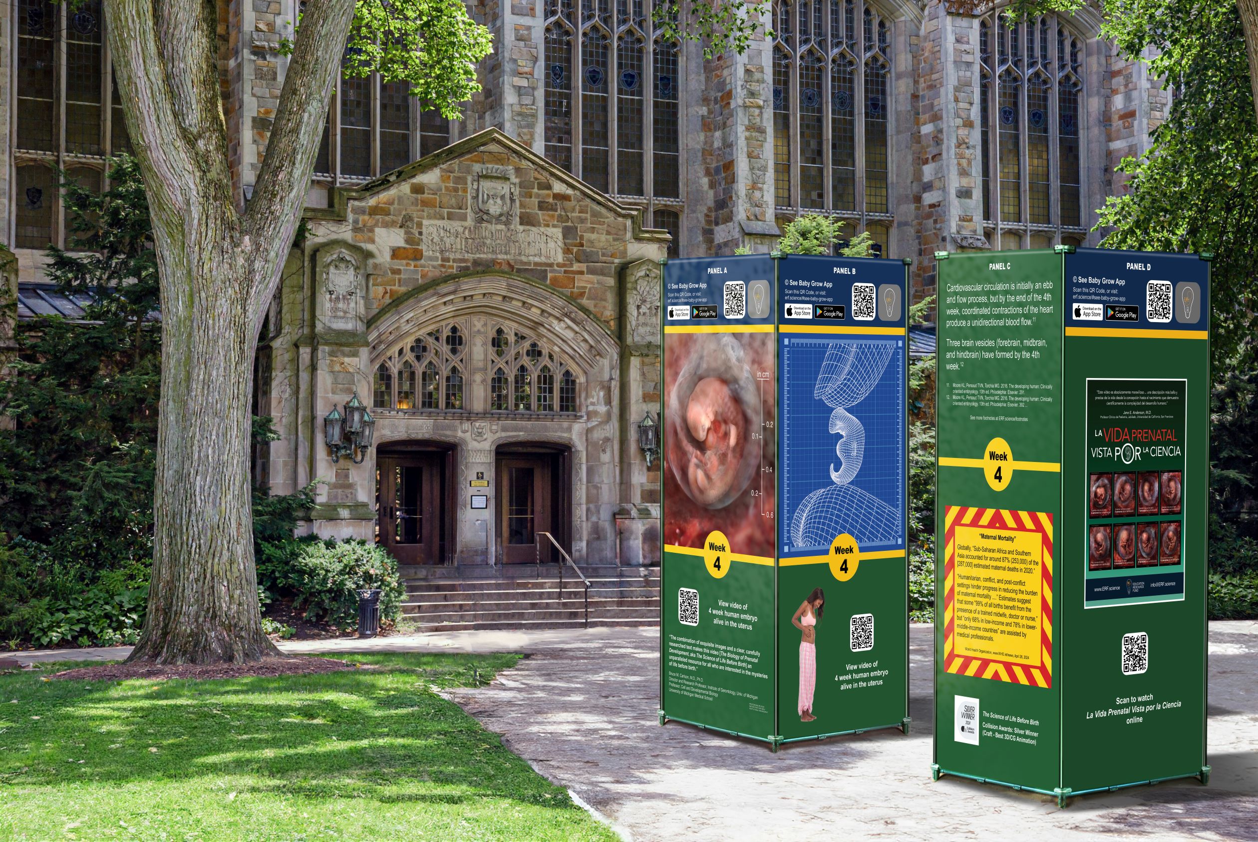
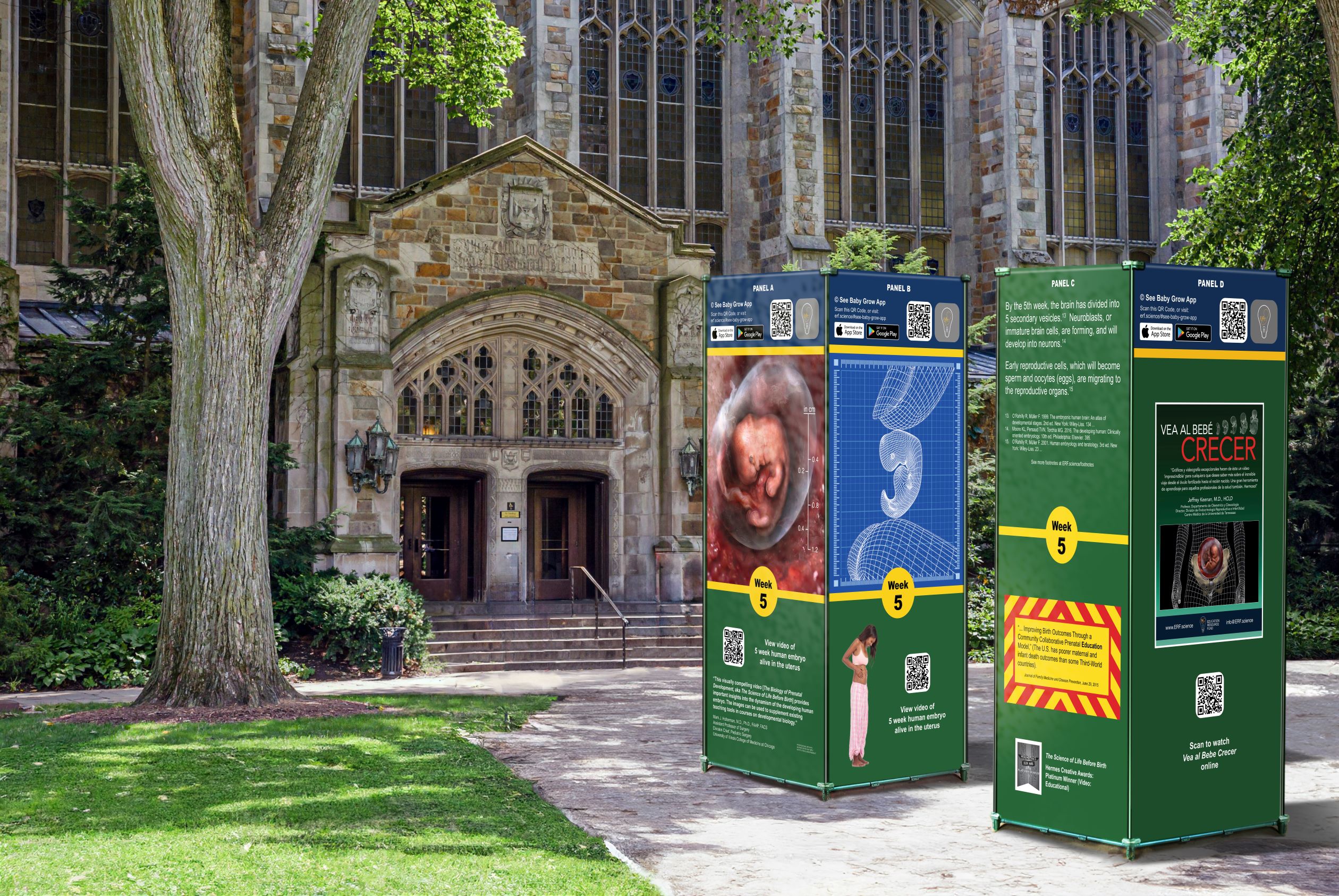
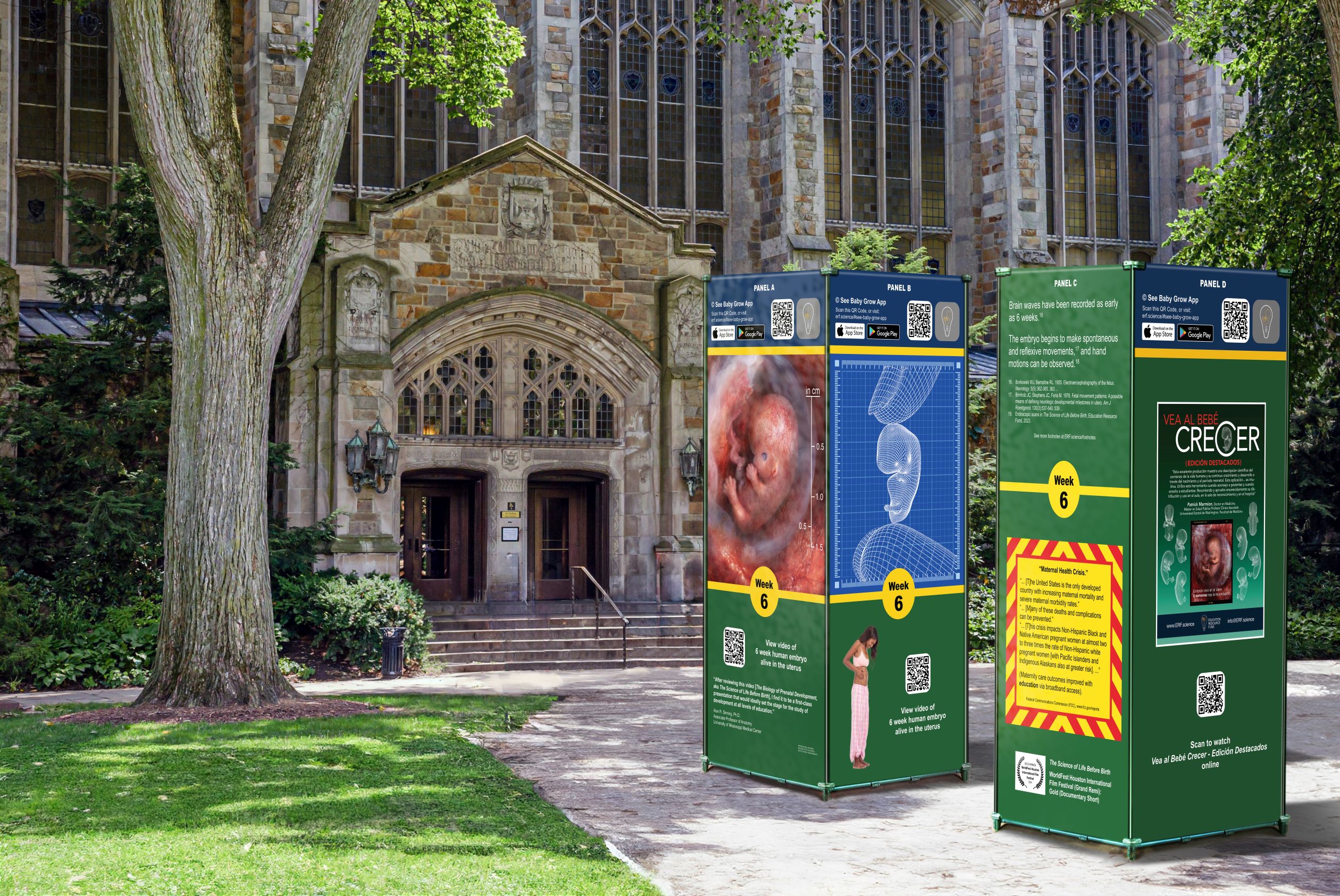
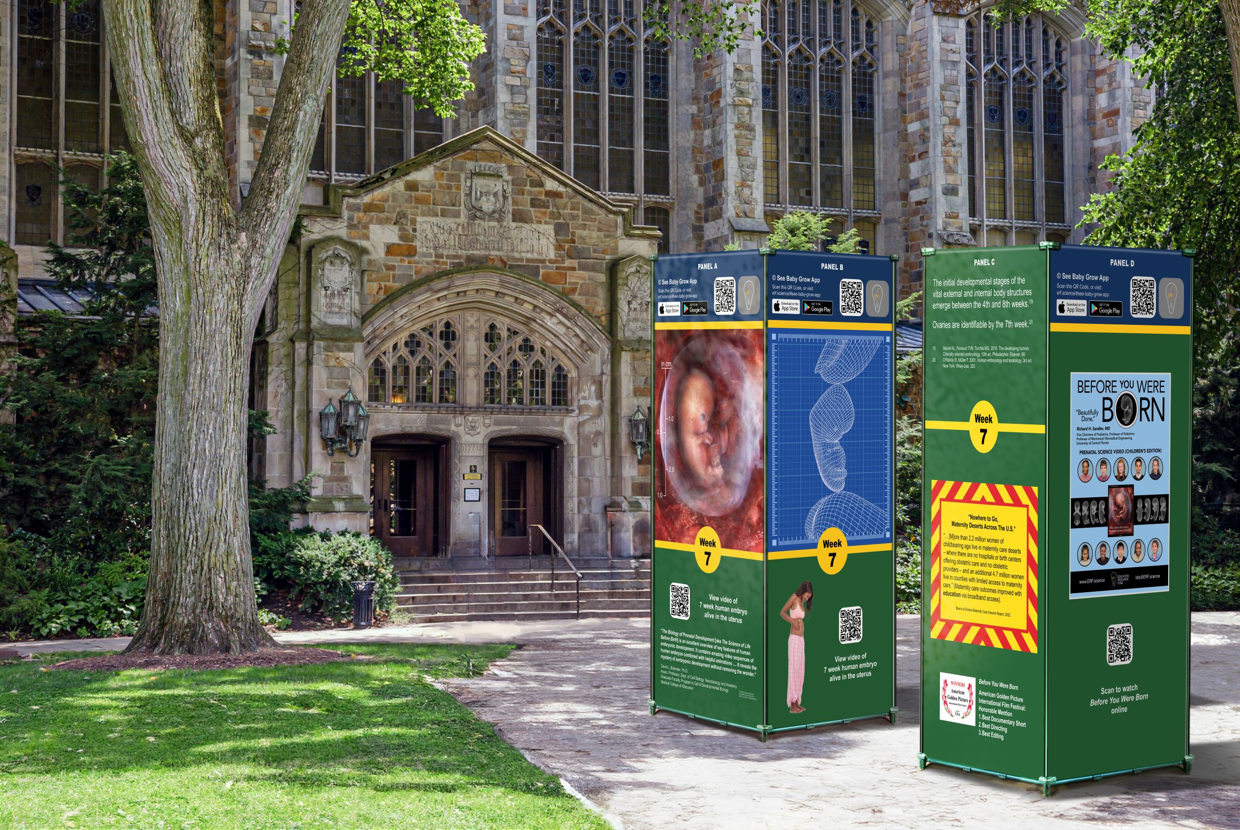
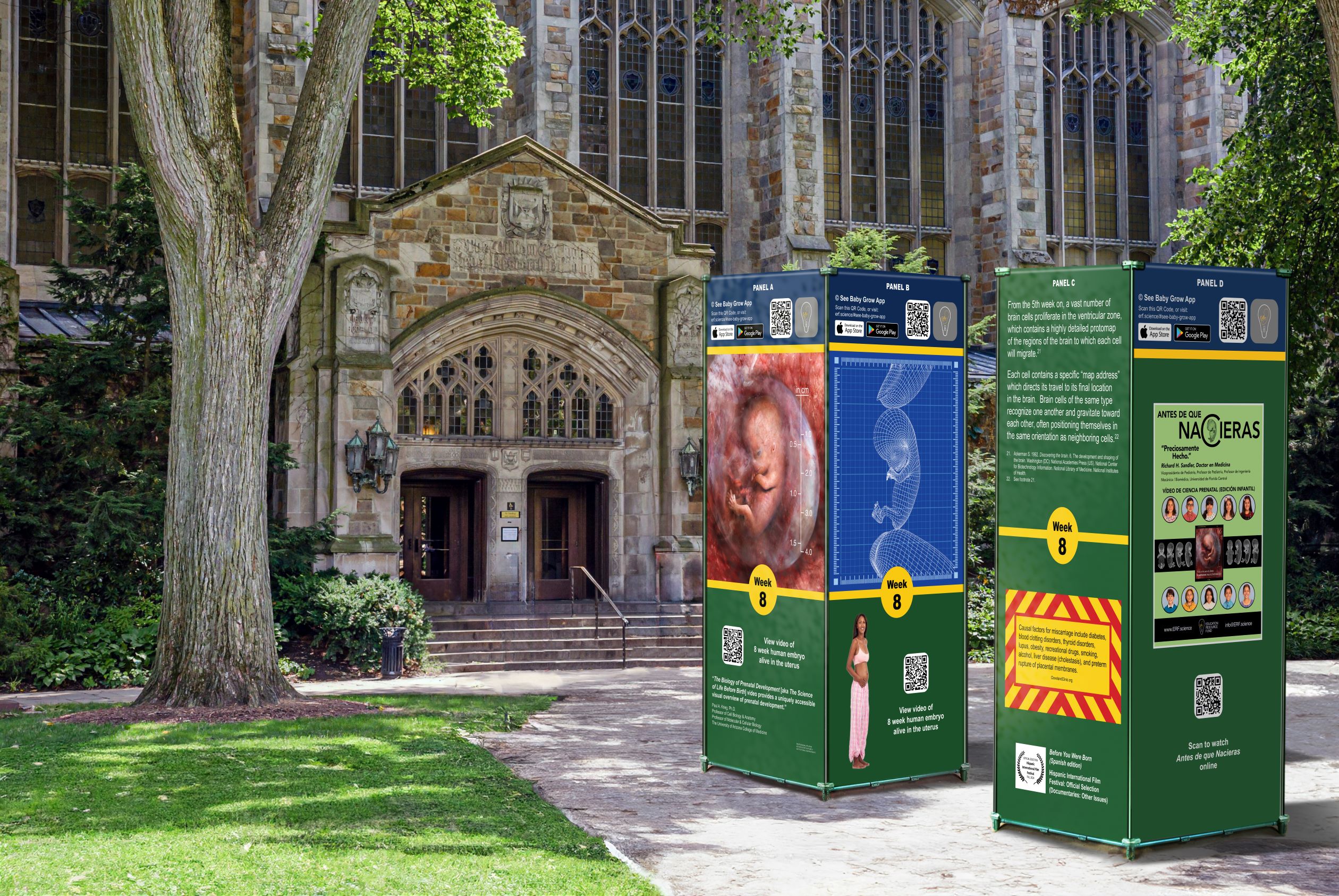
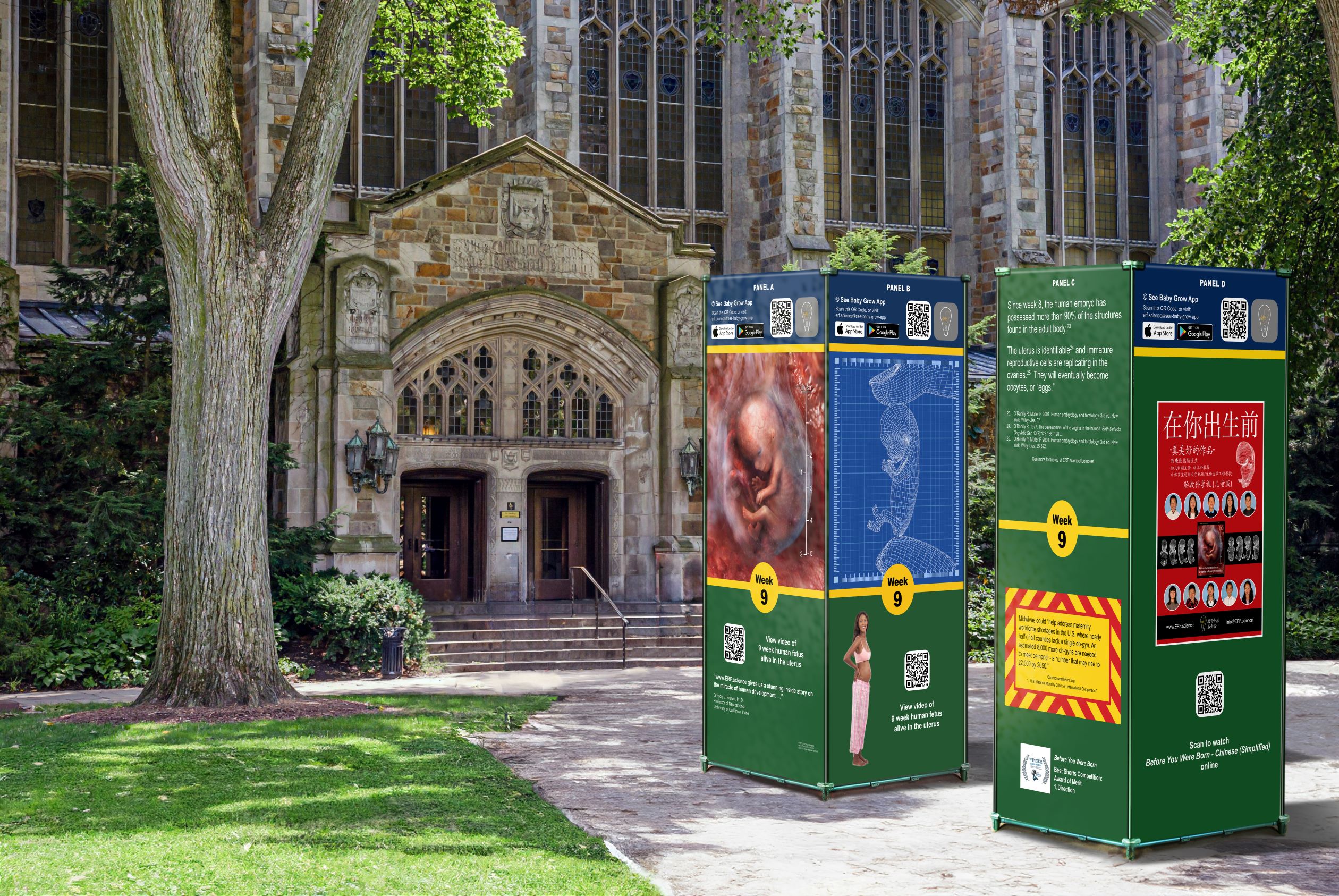
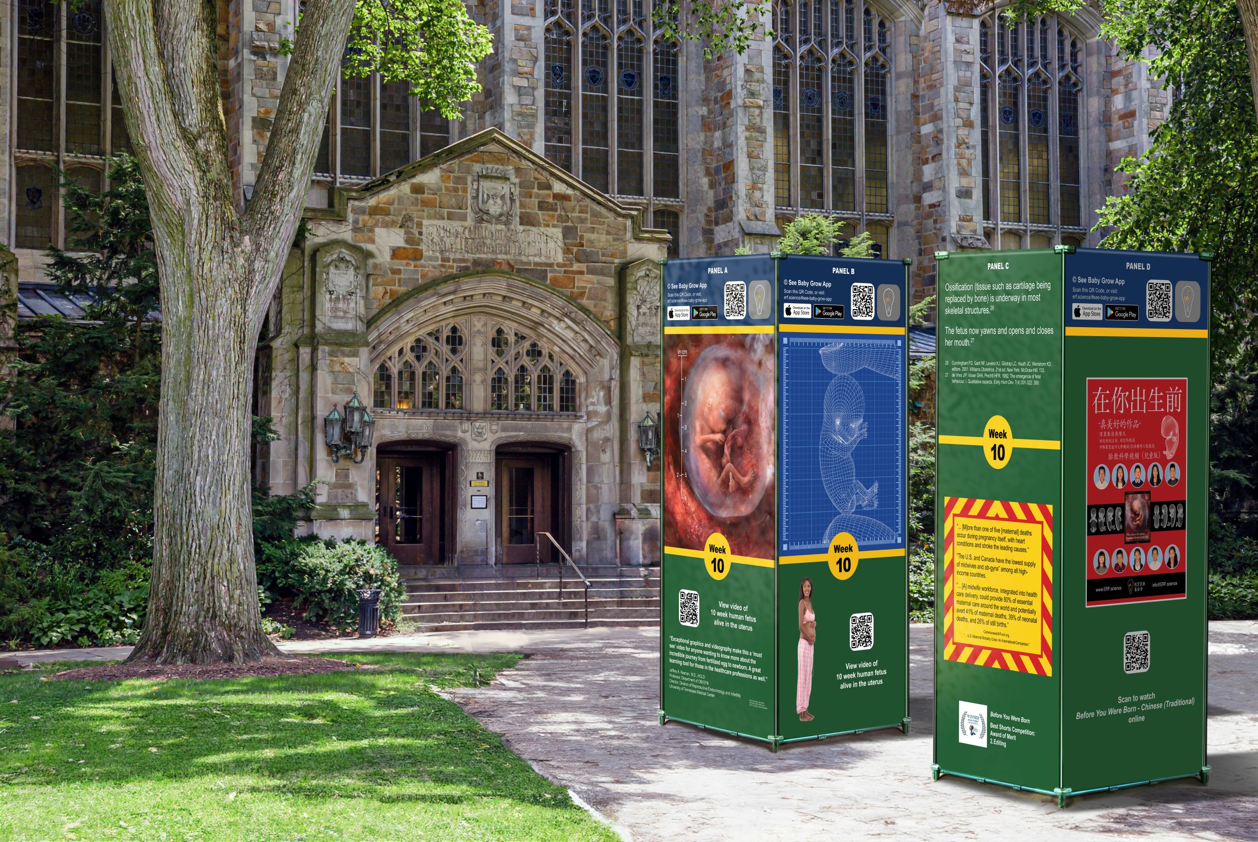
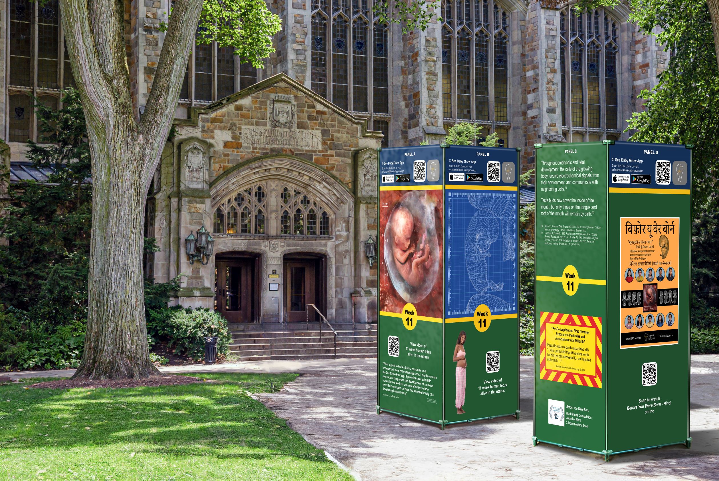
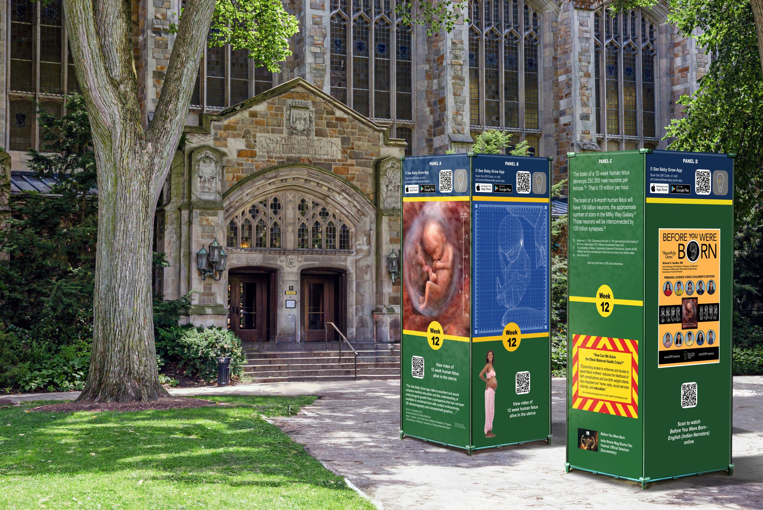
Please see below a comprehensive video archive containing nearly 22 hours of prenatal, endoscopic medical scans. Each scan depicts never-before-seen human embryos and fetuses, alive in the uterus, as they progress through each stage of prenatal development. The image bank will feature a searchable index listing approximately 6,000 of the anatomic structures and/or systems viewable in the video archive. Each search term will be linked to the endoscopic video clip which images its corresponding structure and/or system, chronologically ordered by weeks following fertilization. The program is intended as a reference resource for use by clinicians, academics, researchers, medical and nursing students, as well as students in the health sciences at undergraduate and graduate levels of study. The architecture of this interactive system is designed to accommodate continuous expansion.
Award-winning pregnancy tracker:


2024 AVA Digital Awards
Platinum Winner
Public Service Announcements


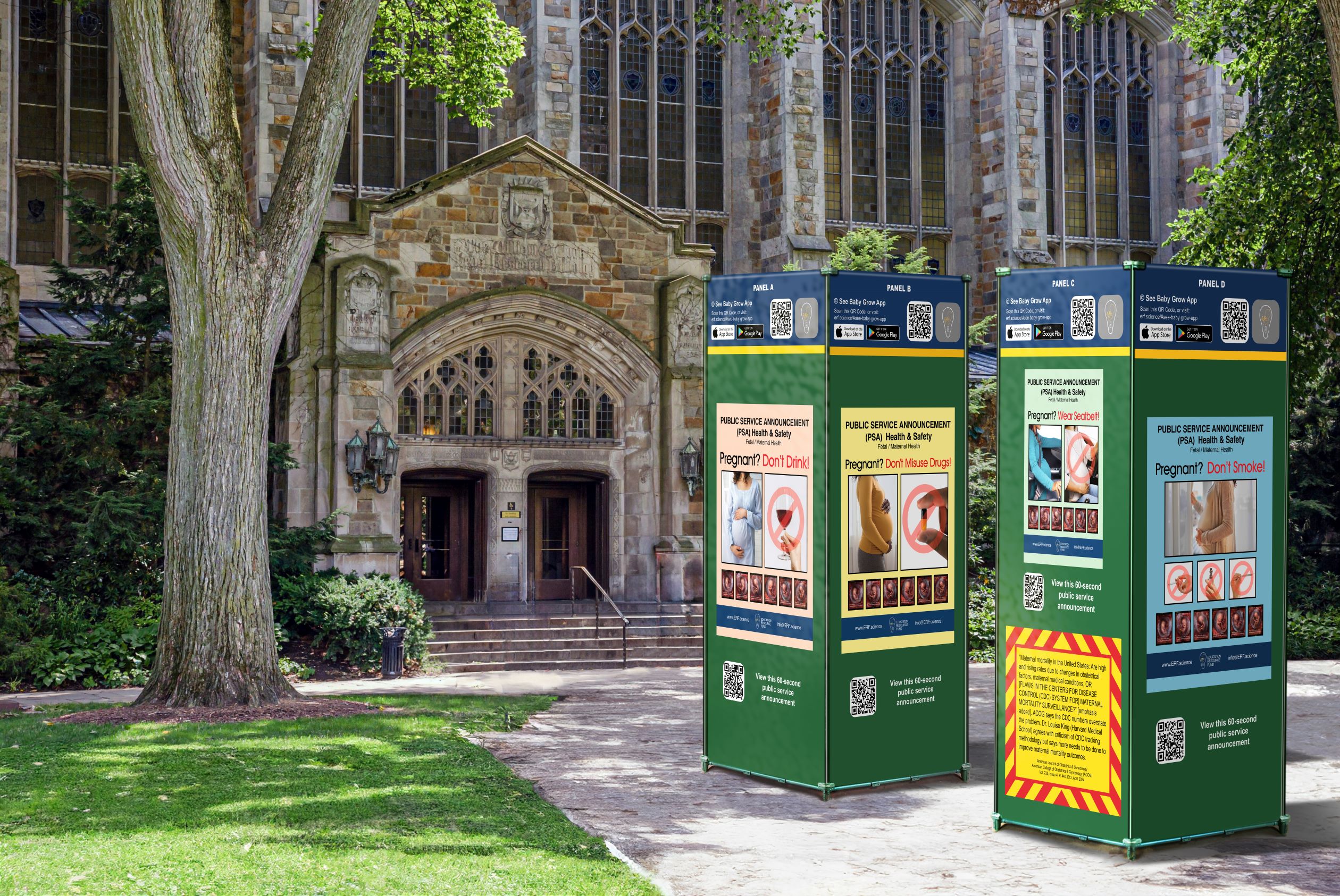
Pregnant? Don’t Smoke!
Pregnant? Don’t Drink!
Pregnant? Don’t Misuse Drugs
Pregnant? Wear Seatbelt!
Science Documentary Films
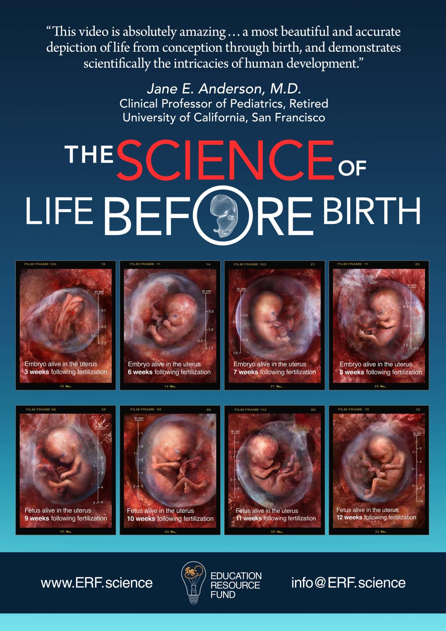
The Science of Life Before Birth
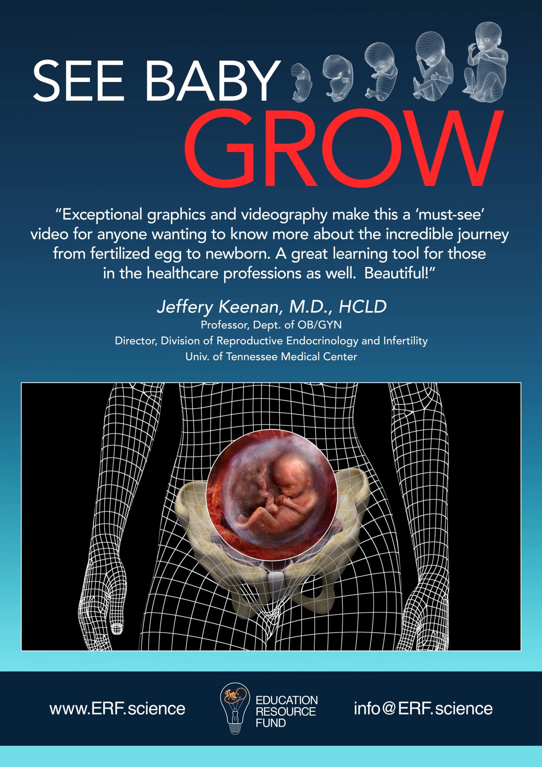
See Baby Grow
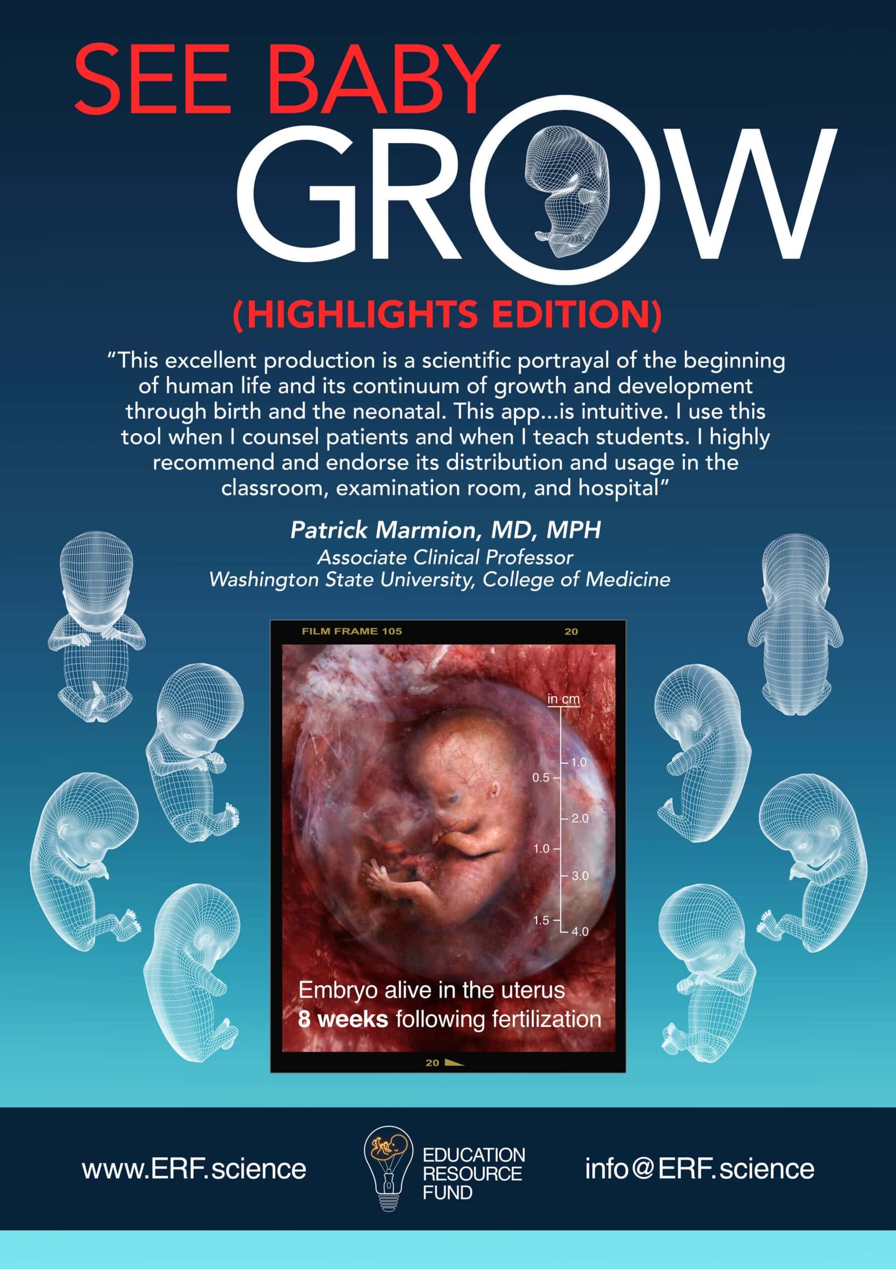
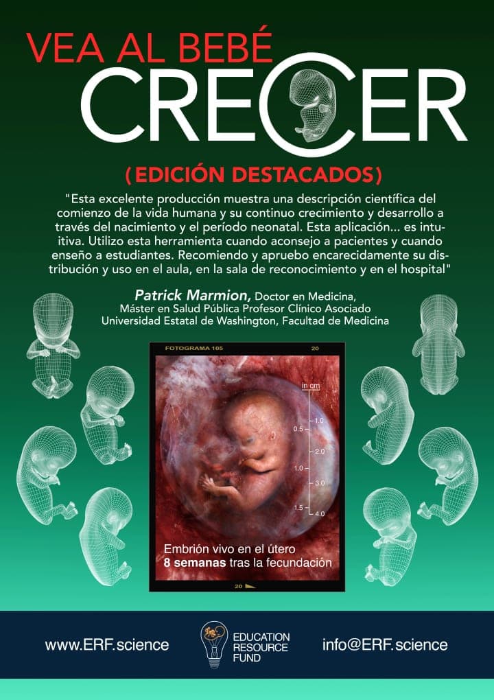
See Baby Grow (Highlights Edition)
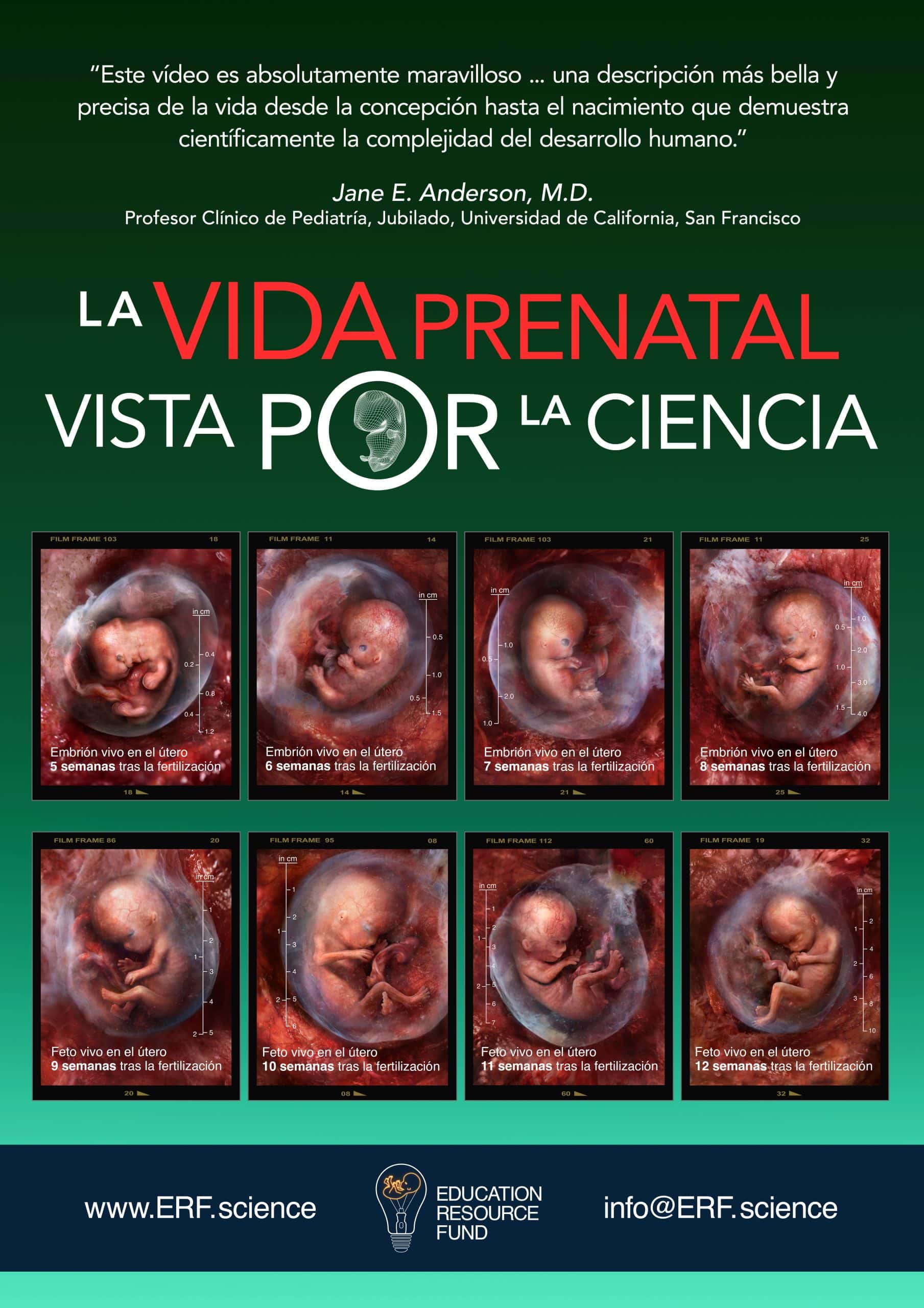
The Science of Life Before Birth – Spanish
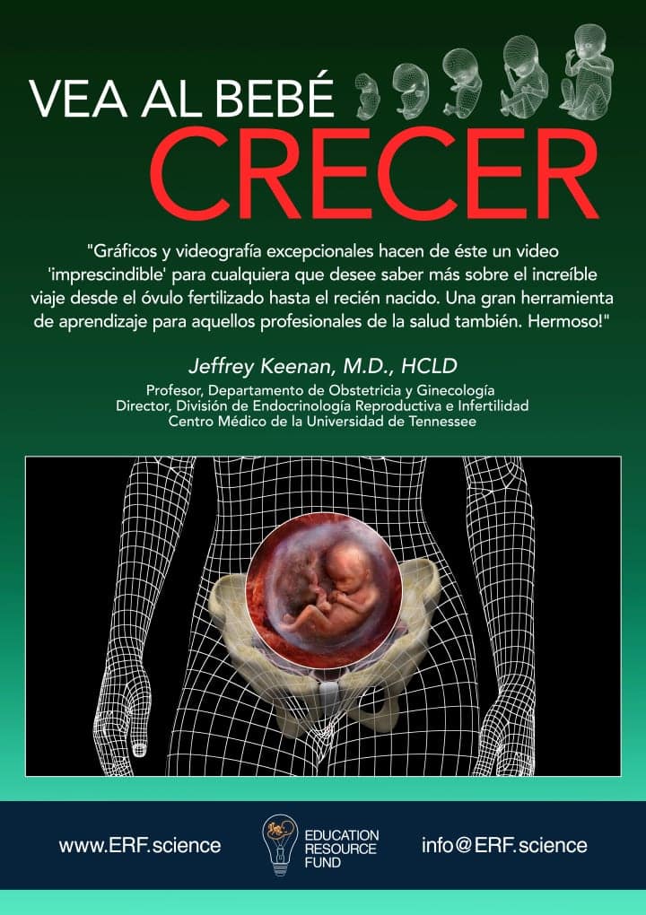
See Baby Grow – Spanish
See Baby Grow (Highlights Edition) – Spanish
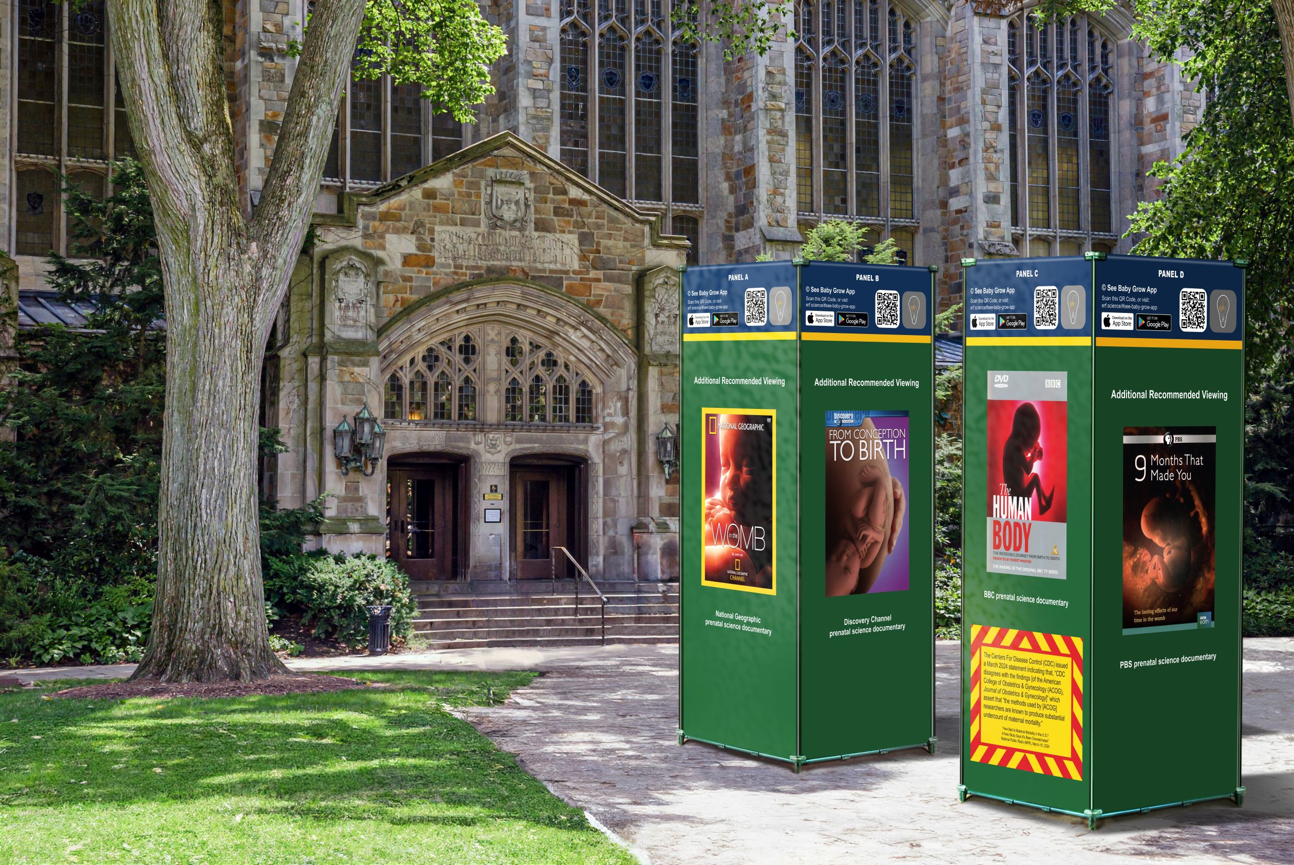
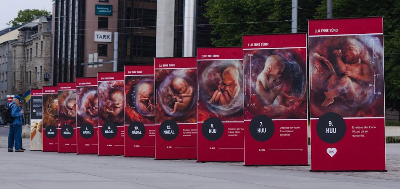
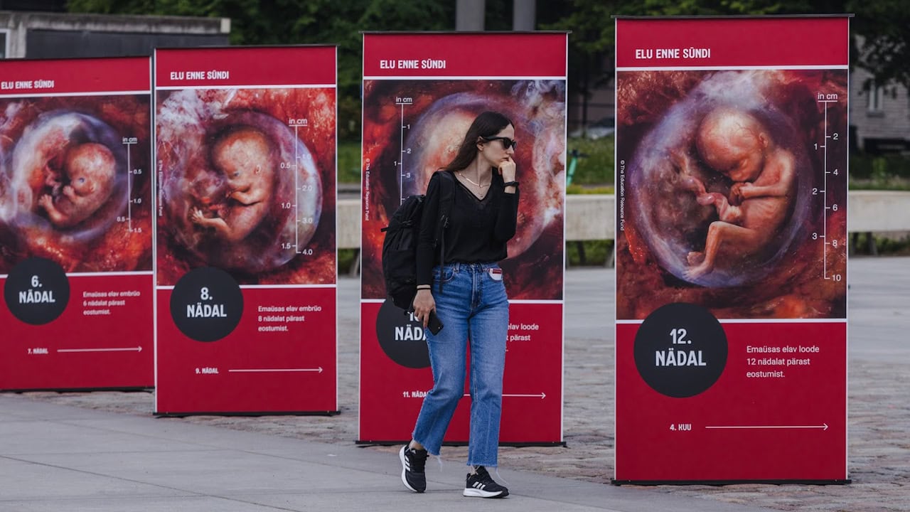
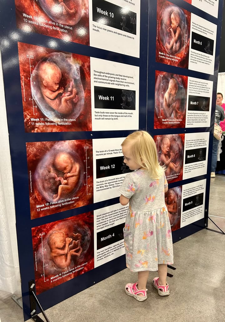
Contact [email protected] for information related to pricing and availability of maternal-fetal health exhibits, now offered by the Education Resource Fund (ERF) for purchase or lease. ERF offers educational display panels to qualified non-profit organizations at prices which cover only ERF’s actual production costs and shipping expenses. International inquiries concerning foreign language translations are also invited.
Award-winning prenatal science documentary:


2024 Collision Awards
Silver Winner

2024 Horizon Interactive Awards
Bronze Winner

2024 Atlanta Movie Awards
Semi-Finalist
Physicians (especially OB/GYNs, pediatricians, and family practice doctors), as well as healthcare professionals generally, may wish to download the following video for display on a TV monitor in their waiting rooms, exam rooms, etc. This 44-minute video features rare endoscopic and sonographic embryonic and fetal scans which span the entirety of pregnancy. The film can be programmed for looping on most TVs.
The Education Resource Fund’s award-winning prenatal medical imagery is now available at Shutterstock Images (shutterstock.com, “… the leading global platform for licensing from the most extensive and diverse collection of high-quality 3D models, videos, music, photographs, vectors and illustrations …”) as well as Adobe Images (stock.adobe.com, “… powered by a world-class community of creative professionals …”) and iStock and Getty Images (subscribers) (GettyImages.com, “… the world’s foremost visual experts – capturing, creating and preserving content to elevate visual communications everywhere …”).
Award-winning prenatal science documentary for children:
2025 Communicator Awards
Award of Excellence
2025 IndieFEST Film Awards
Award of Merit, Special Mention
Editing

2025 IndieFEST Film Awards
Award of Merit
Documentary Short, Direction

2025 Director’s Cut International Film Festival
Semi-Finalist
Best Documentary

2024 American Golden Picture International Film Festival
Honorable Mention
Documentary Short,
Directing, Editing

2024 MLC Awards
Nominee
Any Length Documentary

2024 Best Shorts Competition
Award of Merit
Documentary Short,
Direction, Editing
2024 Horizon Interactive Awards
Bronze Winner
Short Film/Documentary

2024 KIDS International Family Film Festival
Official Selection
Screenplay Documentary Short Film North America

2024 Indie Shorts Mag Short Film Festival
Finalist
Documentary
2024 Atlanta Movie Awards
Semi-Finalist
Documentary Short

2025 Melbourne Independent Film Festival
Certificate of Appreciation
Best Film Editing
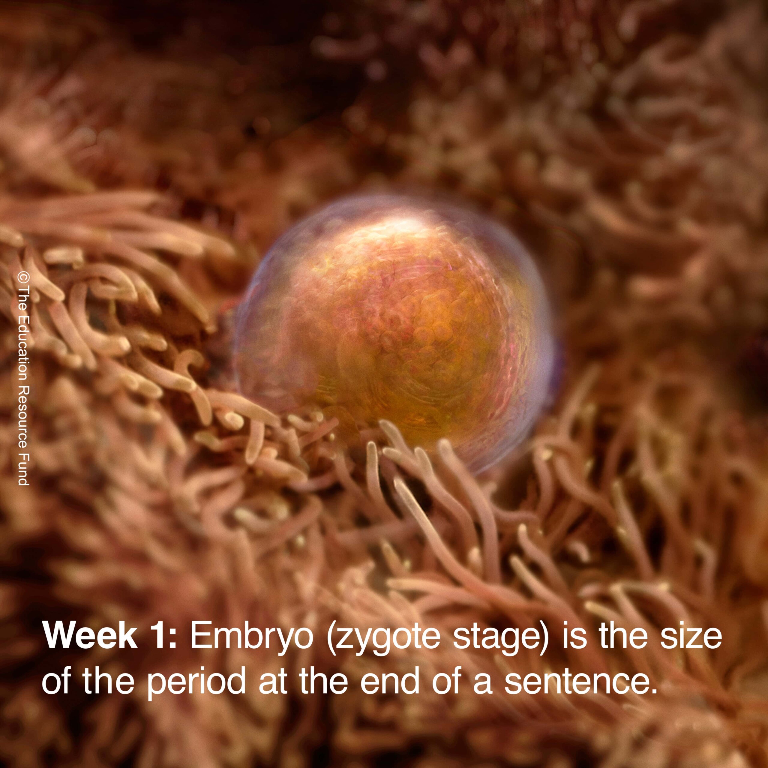
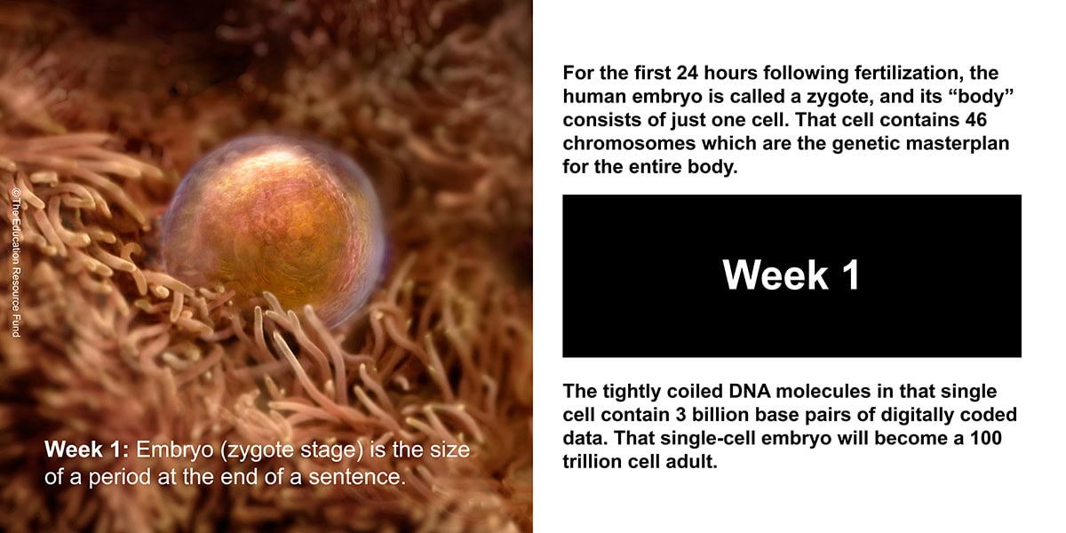
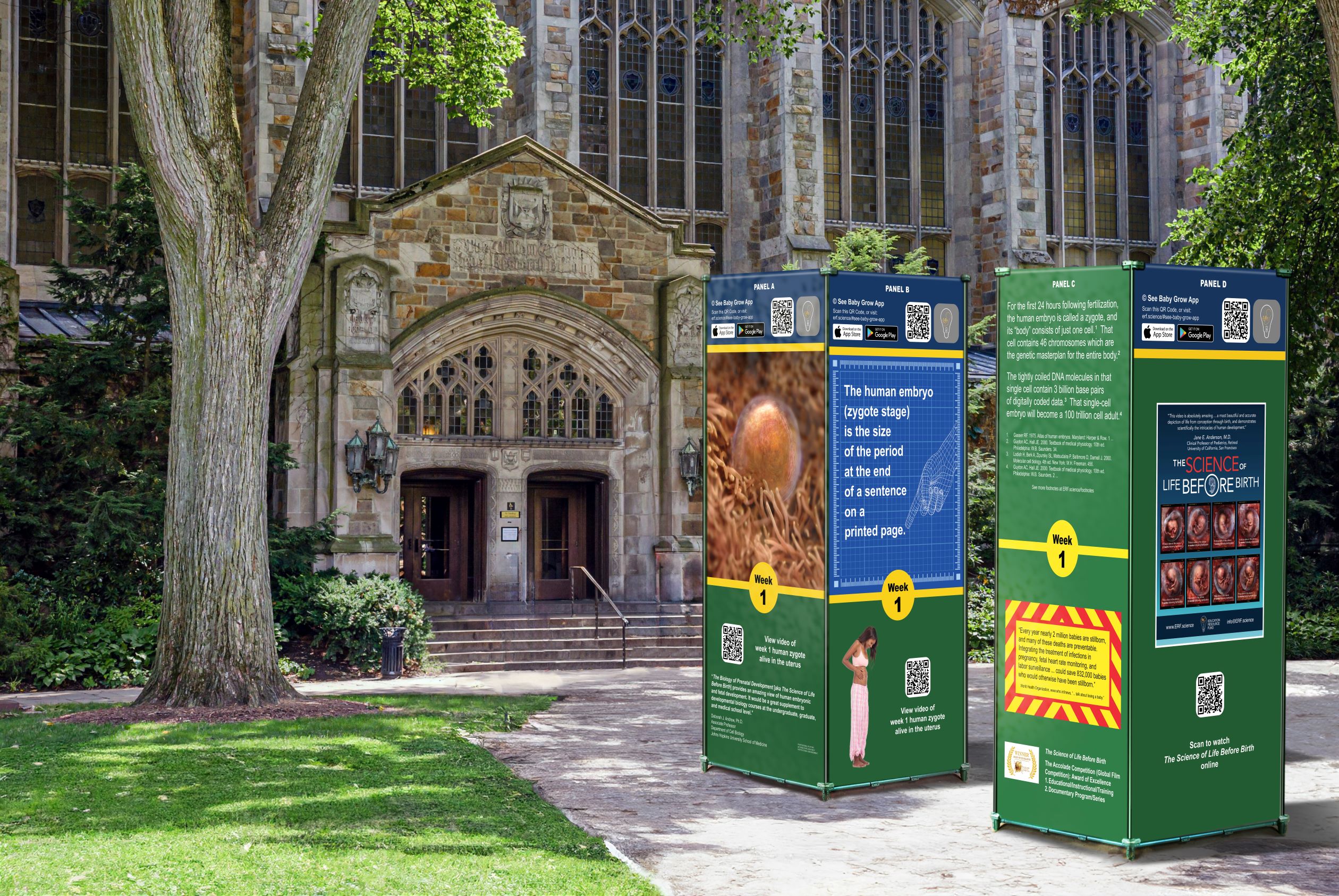
Biologically speaking, “human development begins at fertilization,” when a woman and a man each combine 23 of their own chromosomes through the union of their reproductive cells. The DNA in the 46 chromosomes of the resulting embryo (zygote stage), then only one cell in size, already contains some 3 billion base pairs of digital data, the genetic blueprint for the entire human body. ...
The human heart will beat 3 billion times over the course of an average lifespan.
The human circulatory system contains 20-30 trillion blood cells at any given time.
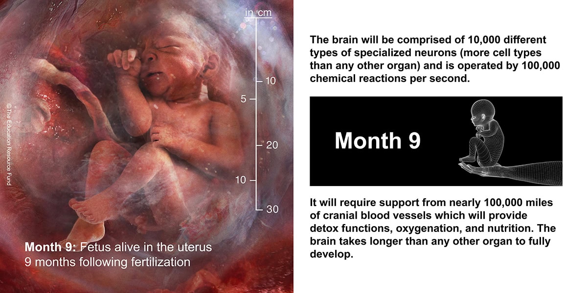
The human brain contains 100 billion neurons.
The neurons in the human brain are linked to one another by 100 trillion synaptic connections.
Award-winning Spanish-language prenatal science documentary for children:

2024 Hispanic International Film Festival
Official Selection
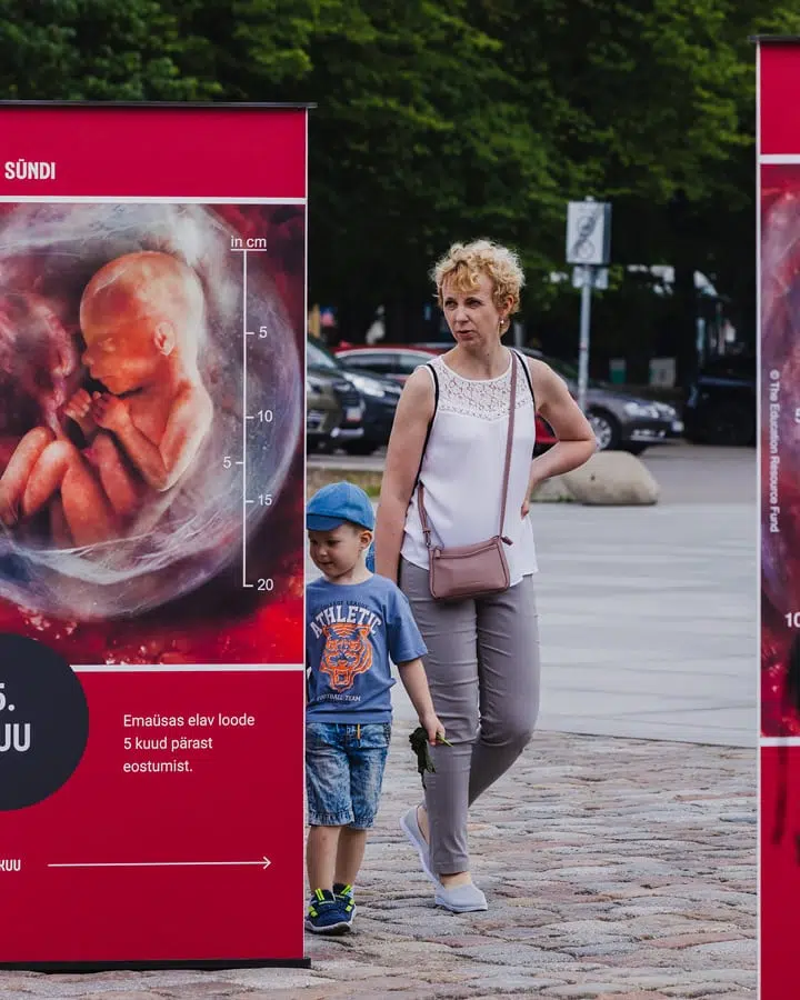
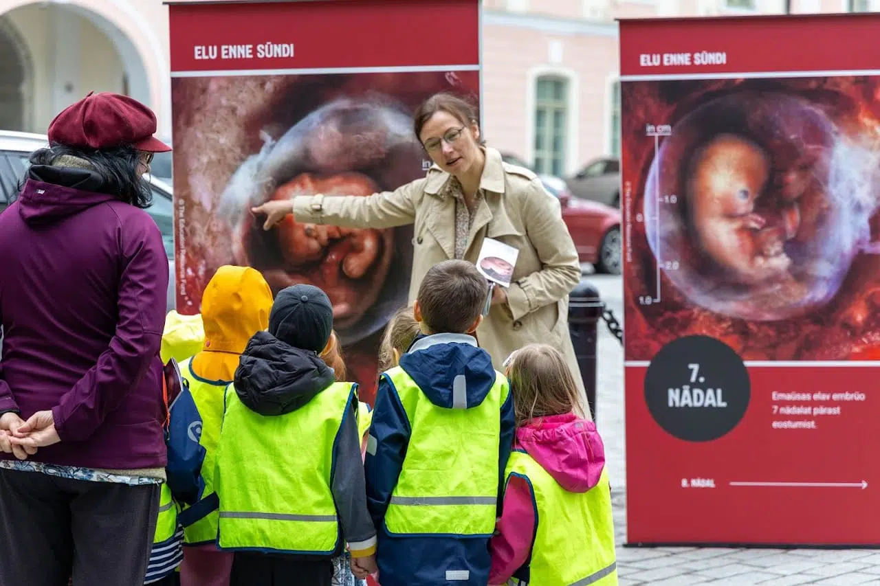
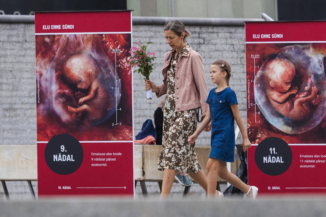
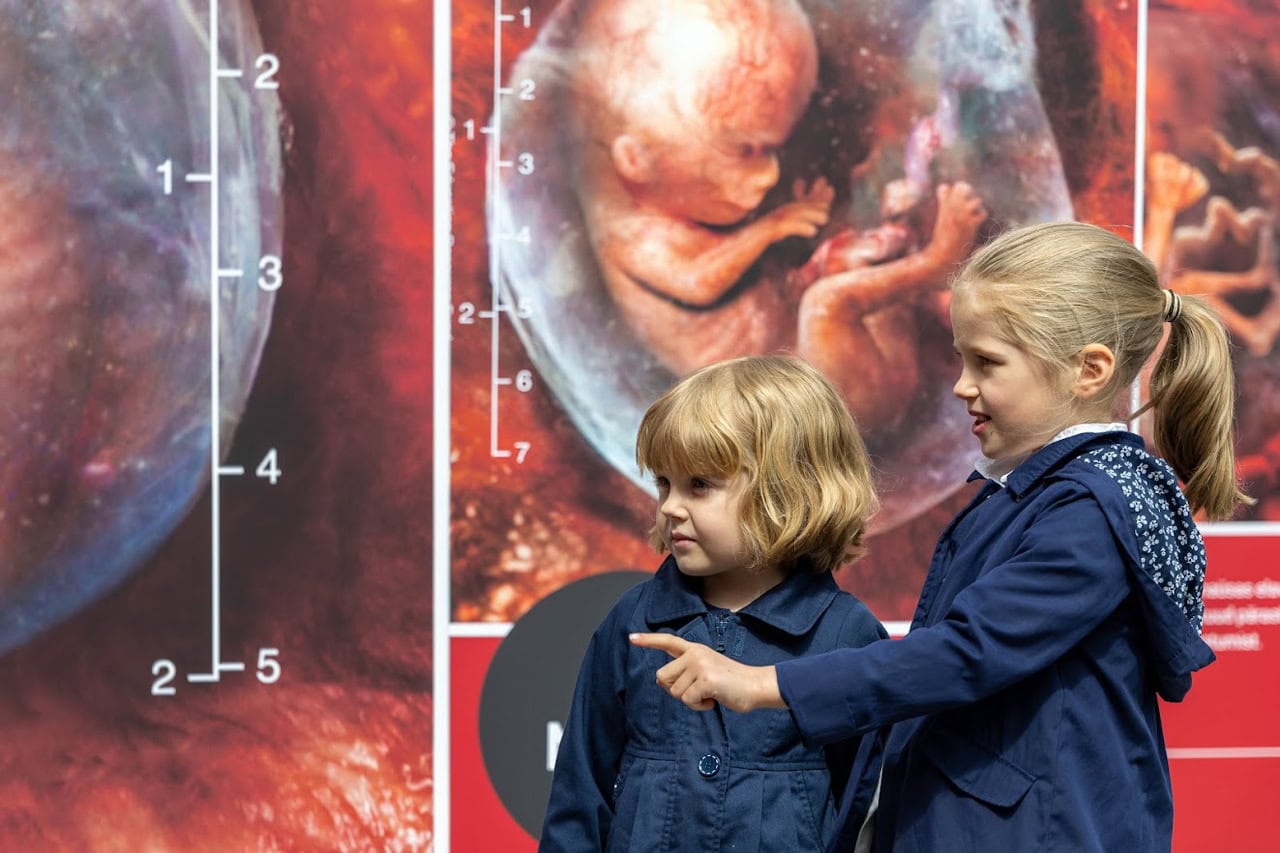
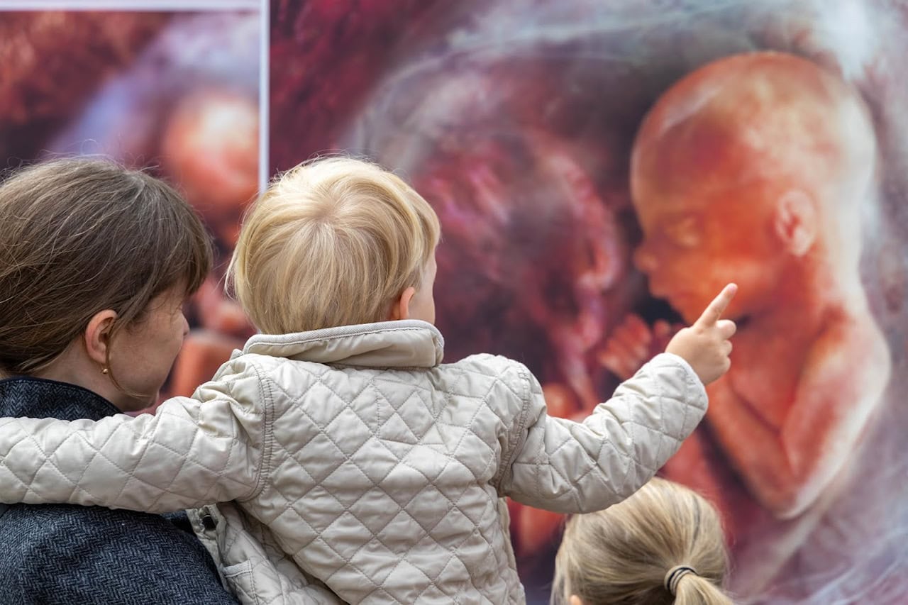
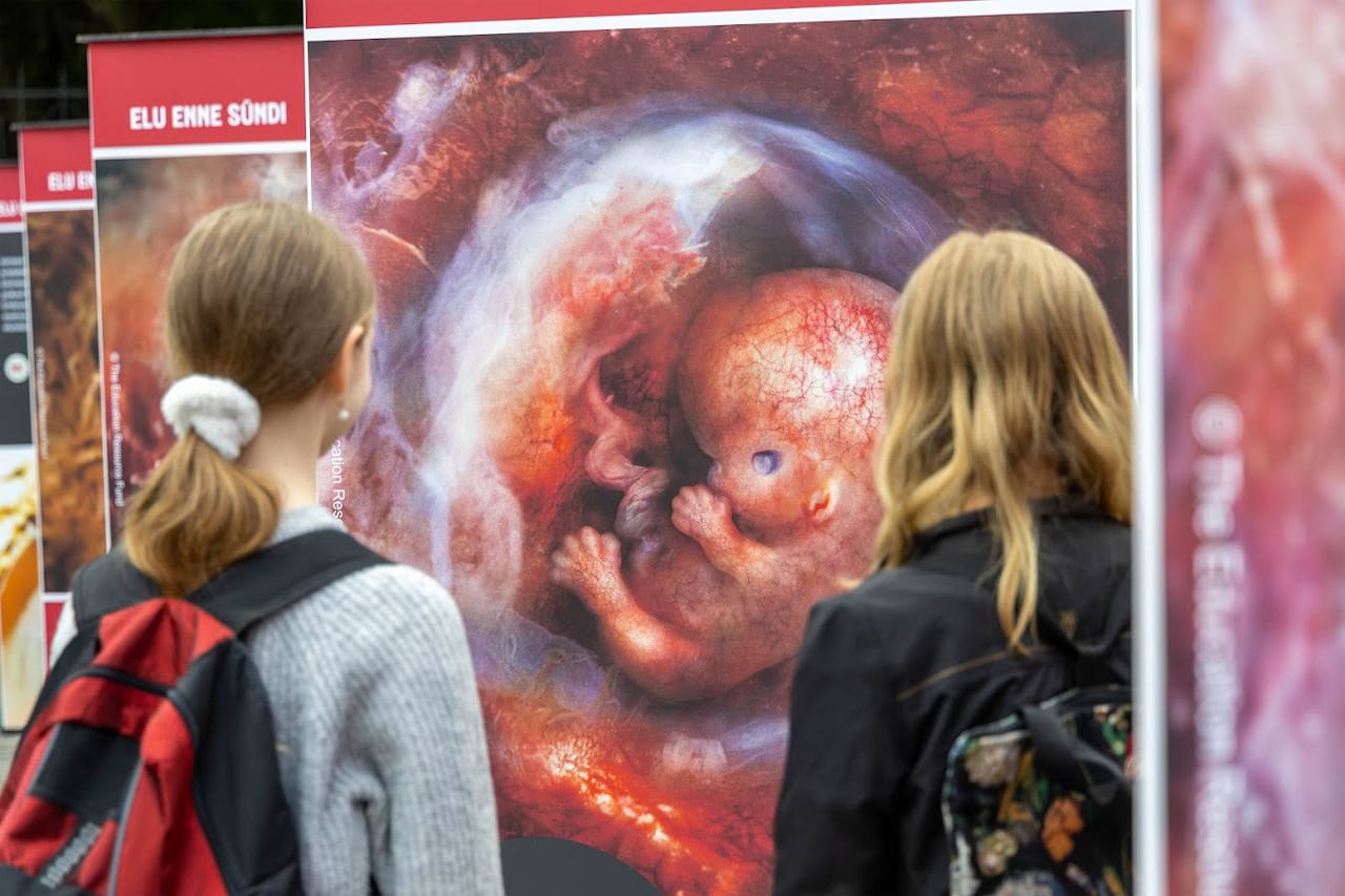
High-resolution images of embryos and fetuses developing in utero


Follow The Science - Fertilization Through 9 Months


Make Science Fun For Kids


Children's Science Documentaries
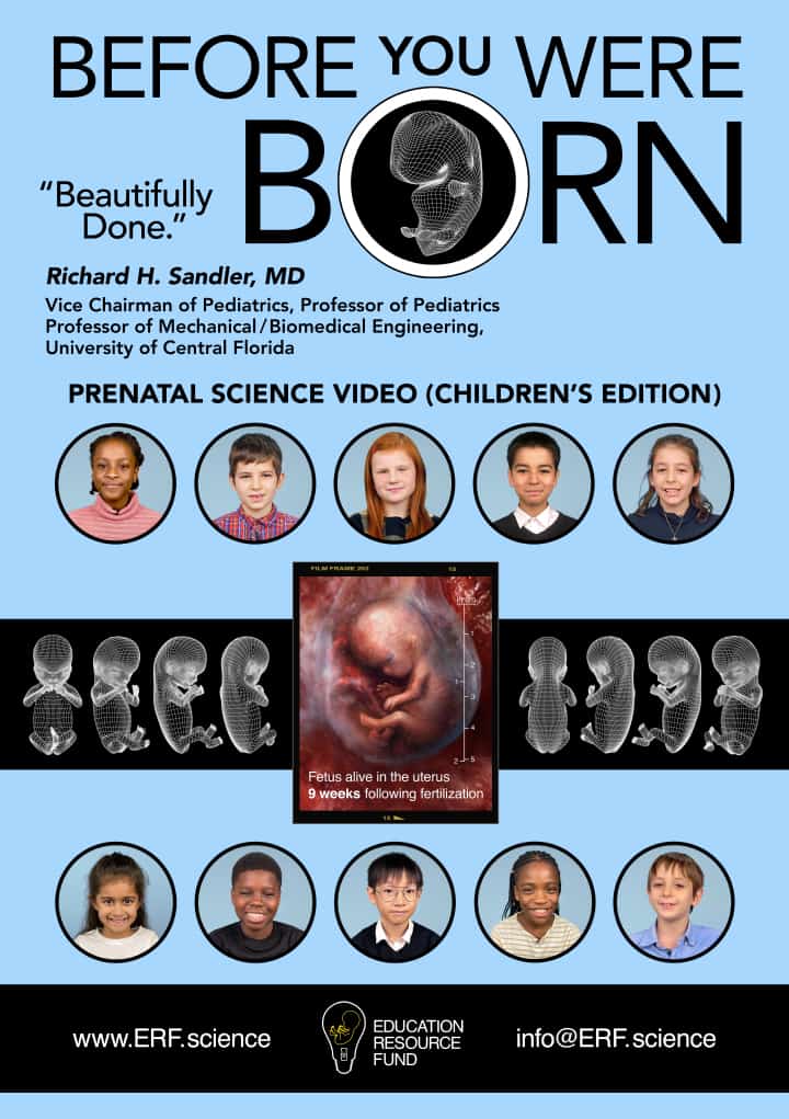
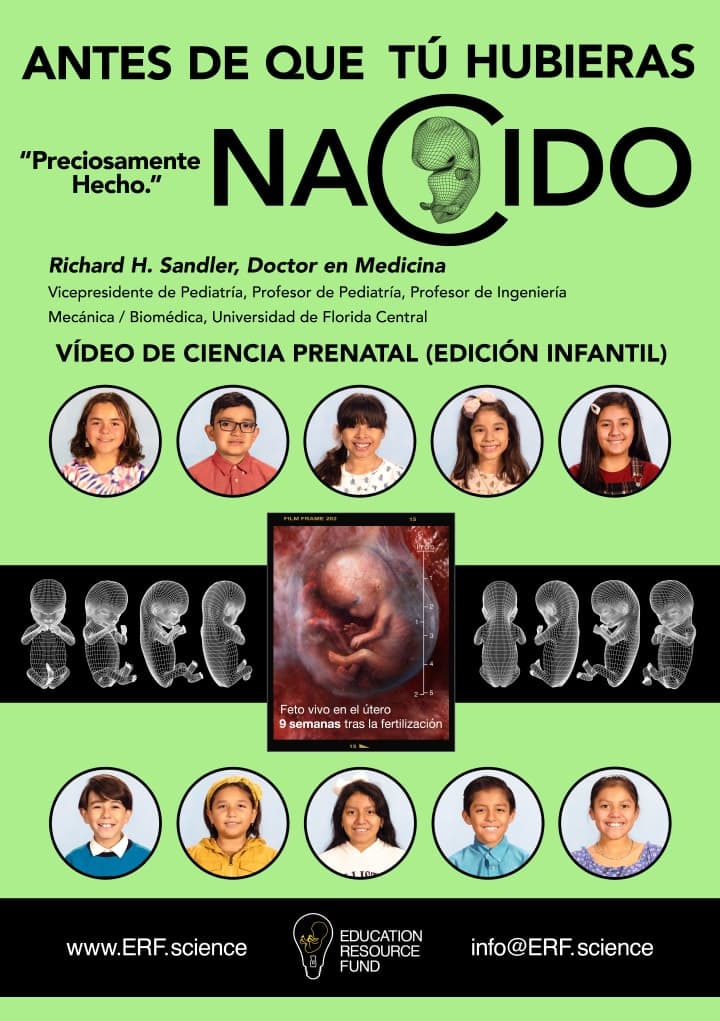


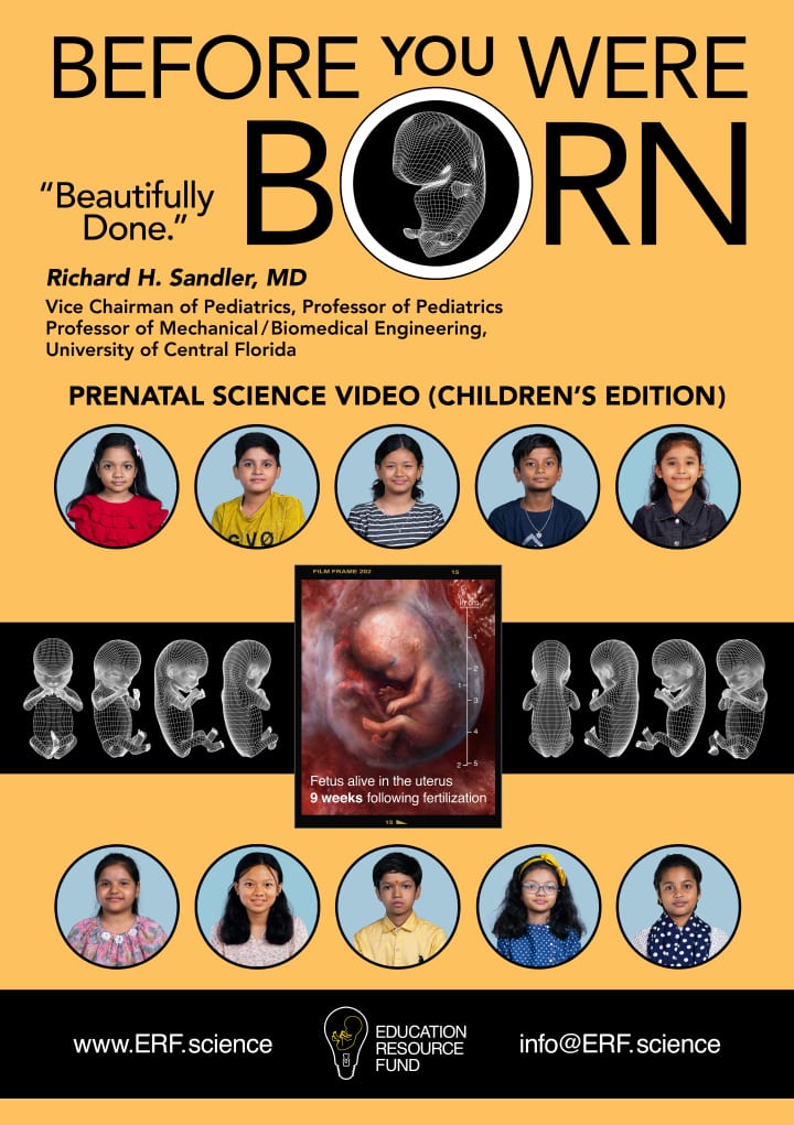
Before You Were Born
Before You Were Born – Spanish
Before You Were Born – Chinese (Simplified)
Before You Were Born – Chinese (Traditional)

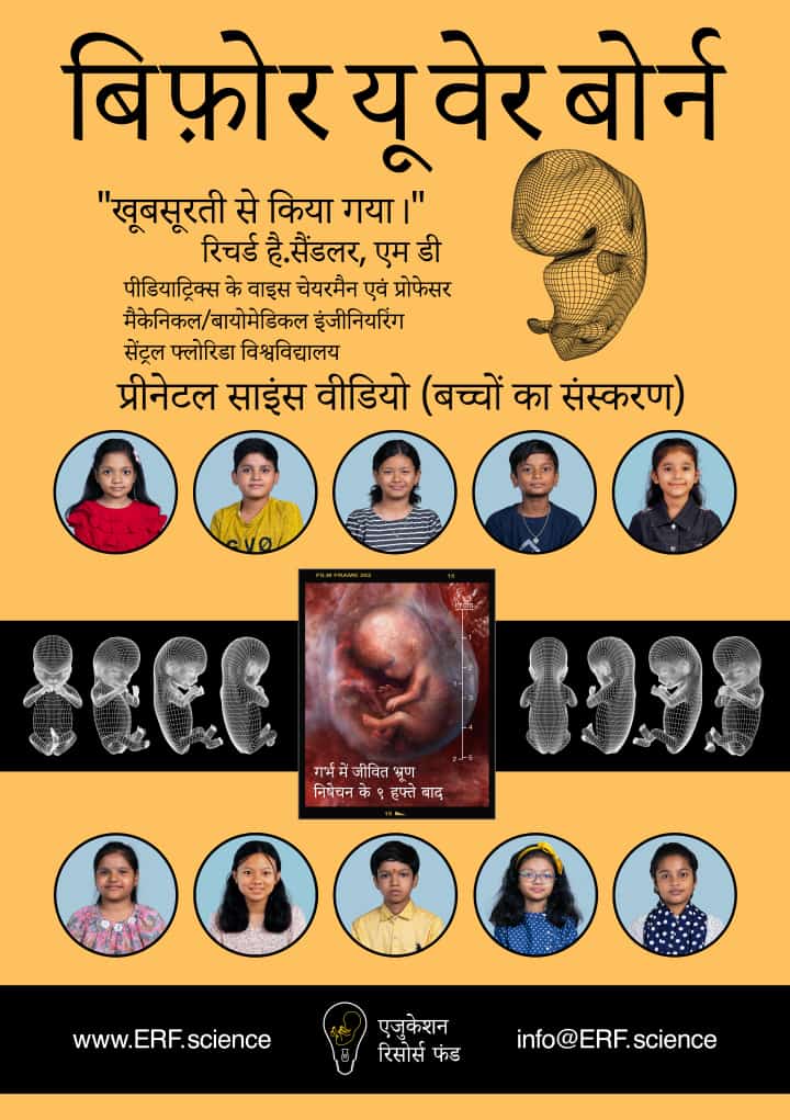
Before You Were Born – Hindi
Before You Were Born – English (Indian Narrators)
COLORING BOOKS
Color using your finger on a phone, or your finger or stylus on a tablet, or your mouse and cursor on a computer, or your crayons on physical pages you print out on paper.


Auto-fill zone coloring
Color with finger or stylus
“I hold a multiple subject teaching credential in the state of California and I have been a public elementary school teacher since 2004. My primary focus has been Kindergarten, First and Second grade. I recently examined the ERF coloring pages which depict embryos and fetuses developing in utero. This is an amazing interactive resource for children of all ages. It’s extremely user friendly. The high-resolution prenatal images next to the coloring book line drawings are fantastic! This is an instructive resource that can be used effectively in the classroom setting. I also reviewed the ERF site where I watched the children’s version of the ERF prenatal video. Like the coloring pages, the children’s edition of the ERF prenatal science documentary is also an amazing resource! All content is age-appropriate for even the youngest children.”
Ellarose Pinkus
Puzzles


Curricular Content


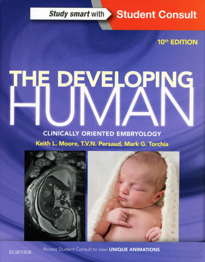
Please see the following Google description (citing Amazon.com book review) of The Developing Human: Clinically Oriented Embryology, the medical textbook from which the Education Resource Fund (ERF) quotes the distinction between the “weeks from last menstrual period” and “weeks from fertilization” embryonic and fetal aging standards:
“The Developing Human: Clinically Oriented Embryology (often referred to as Moore) is widely regarded as a leading, industry-standard textbook in medical education. It has been a core resource since its debut in 1973 and was recently designated as an Essential Purchase for 2025 by Doody’s Core Titles.”
Amazon.com
Excerpt from Moore:
By convention, obstetricians date pregnancy from the presumed first day of the last normal menstrual period (LNMP). This is the gestational age, which in embryology is superfluous because gestation does not begin until fertilization of an oocyte occurs. Embryonic [or fetal] age [also described as fertilization or conceptional age] begins at fertilization, approximately 2 weeks after the LNMP…. The day on which fertilization occurs is the most accurate reference point for estimating [embryonic or fetal] age ….
The Developing Human: Clinically Oriented Embryology. Moore KL, Persaud TVN, Torchia MG. Philadelphia: Elsevier. 10th Ed. 2016. p 87.
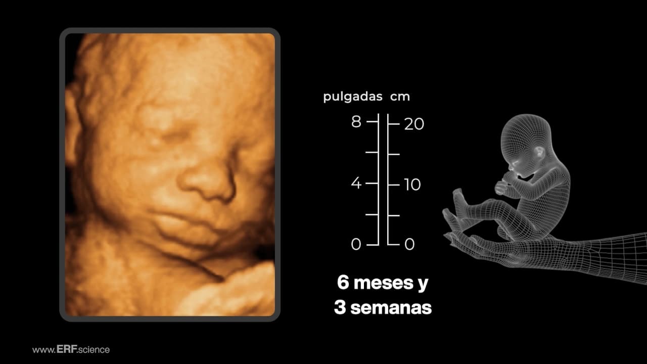
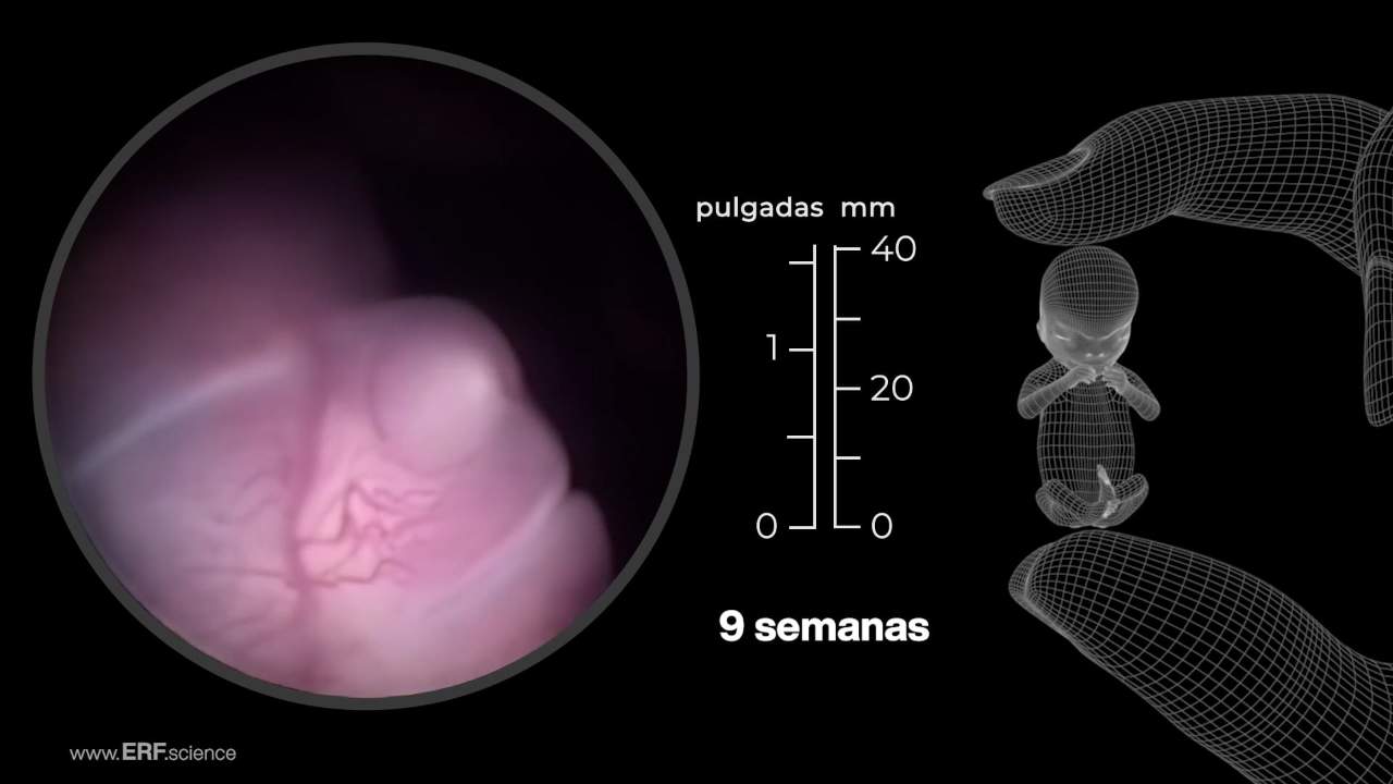
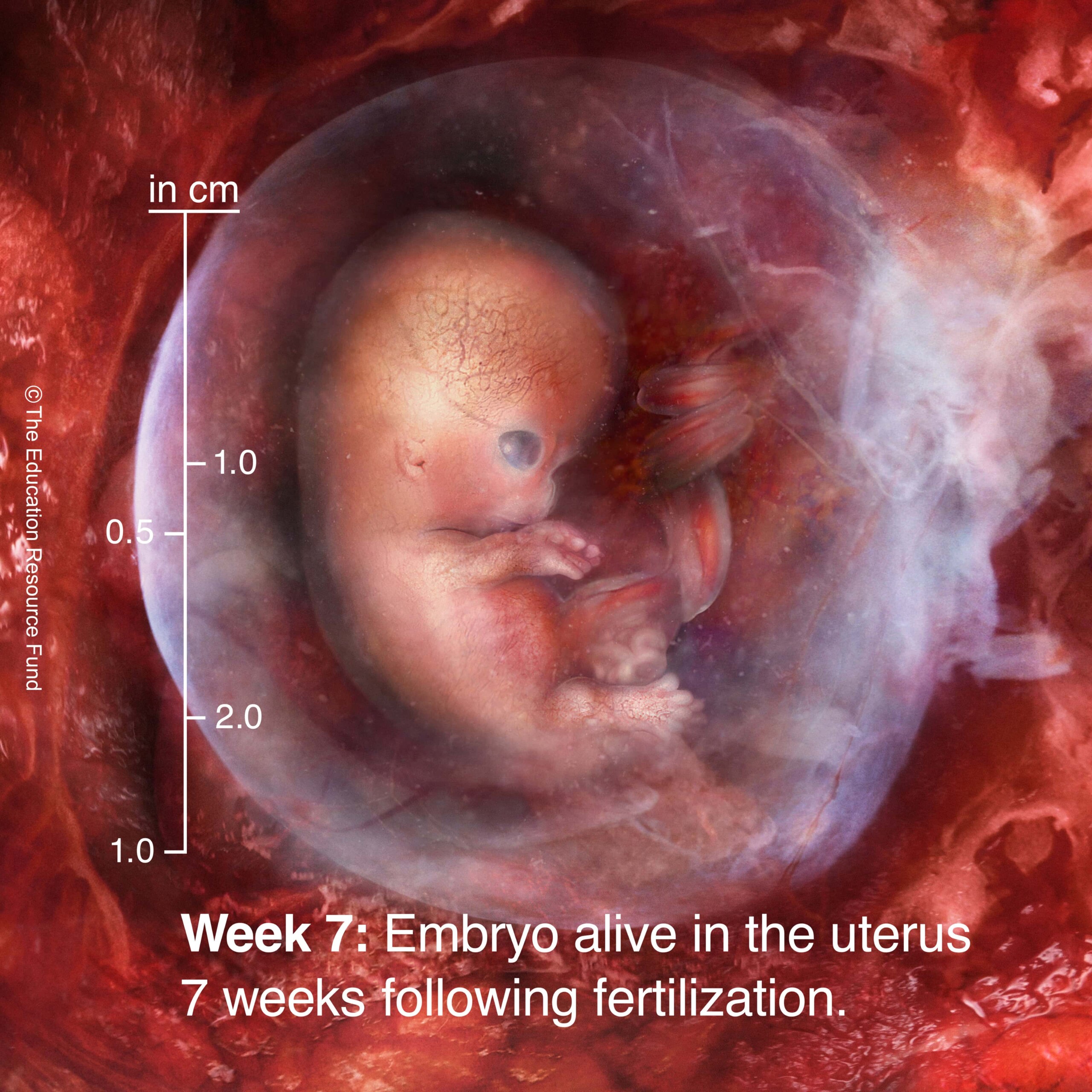
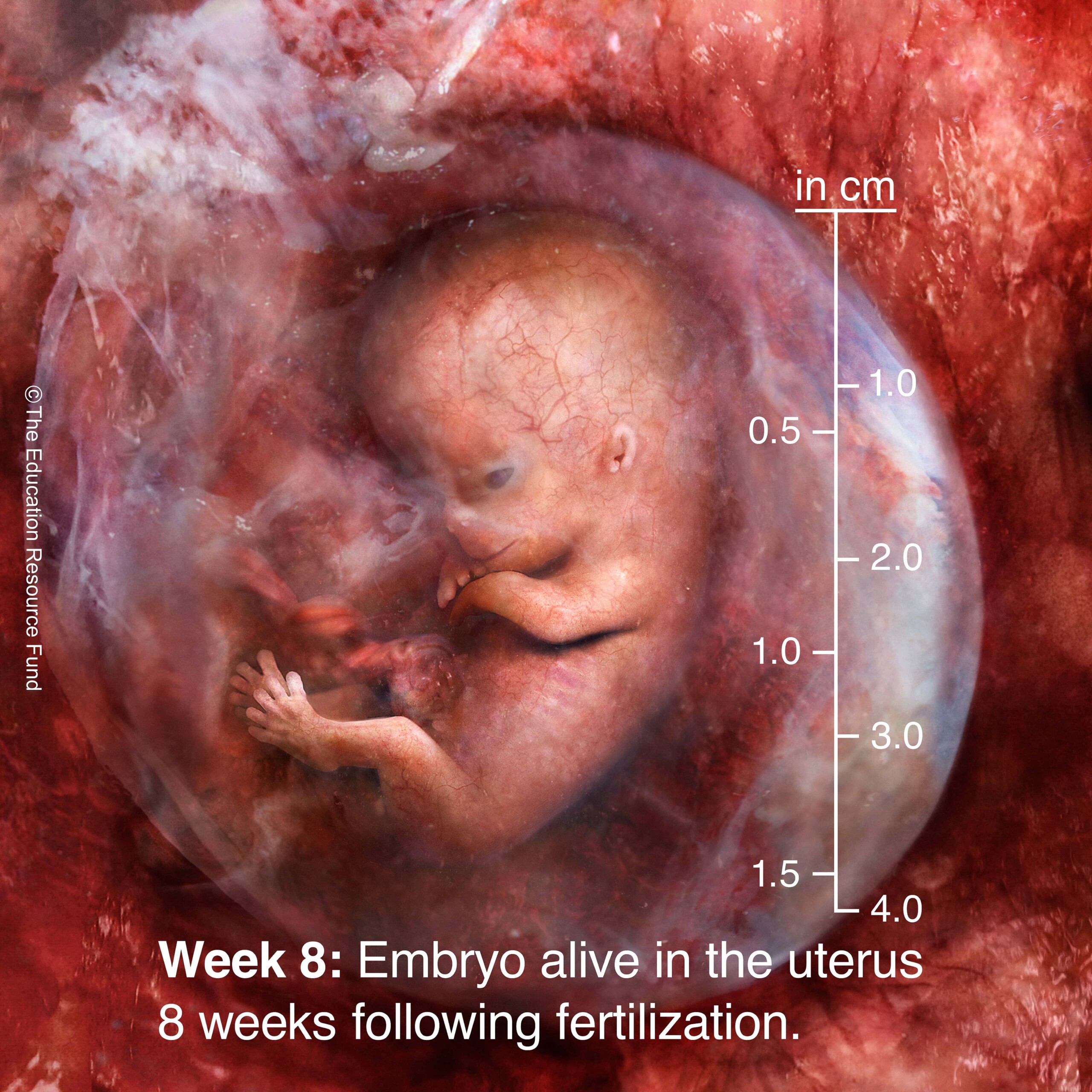
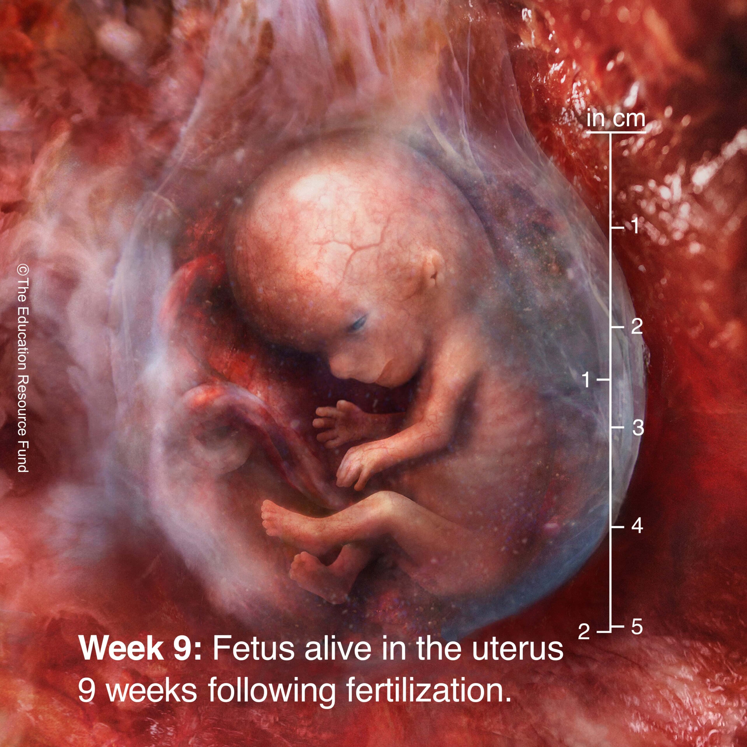
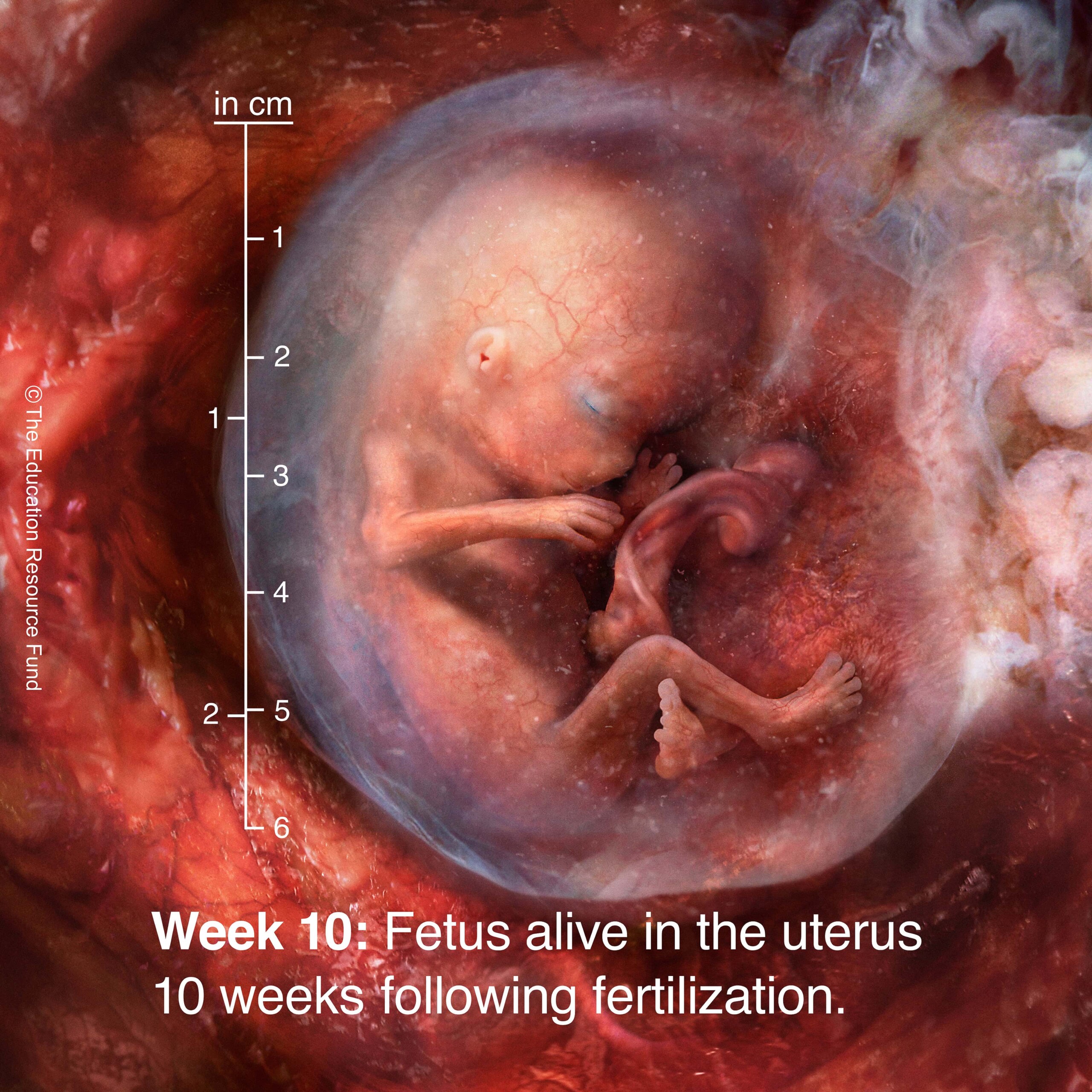
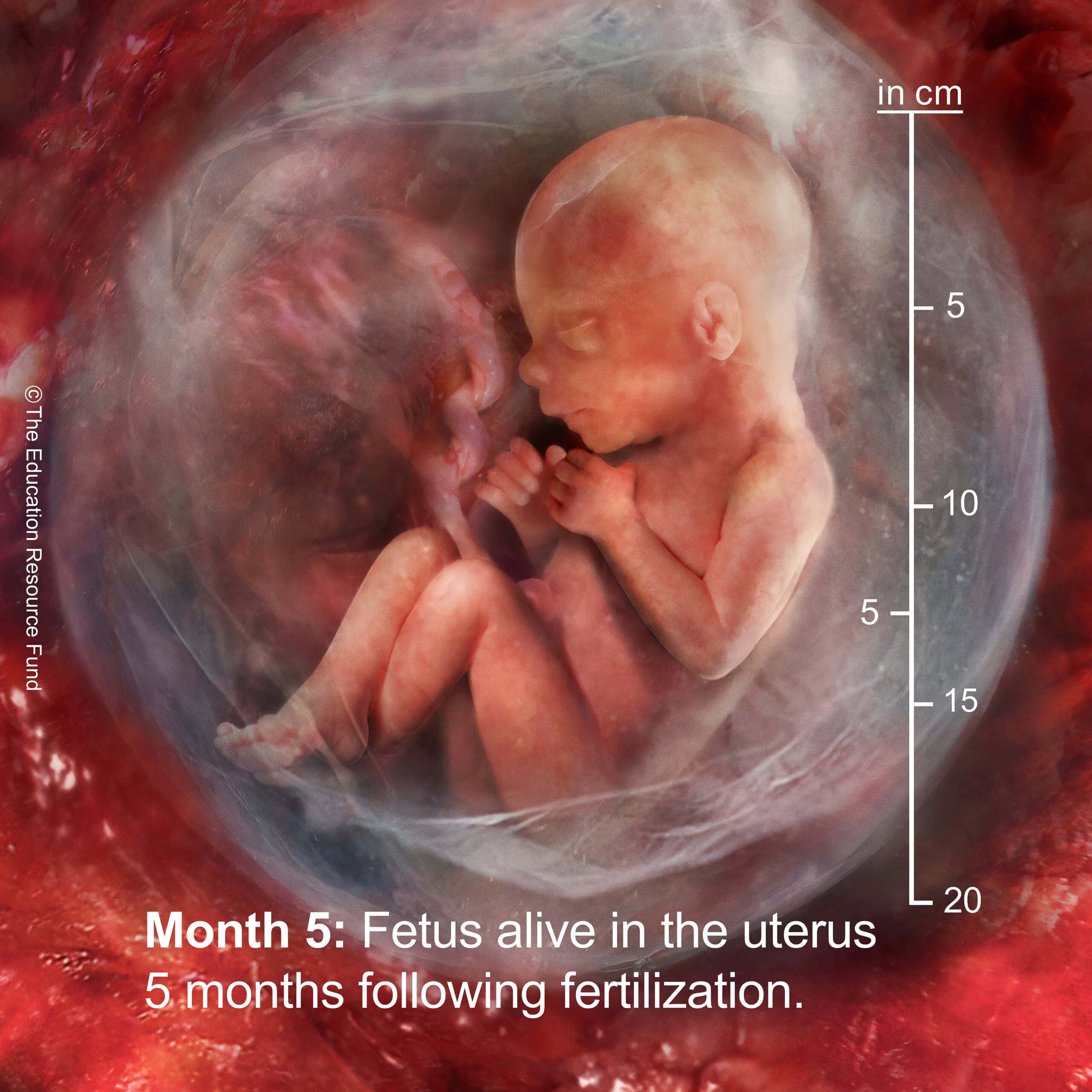
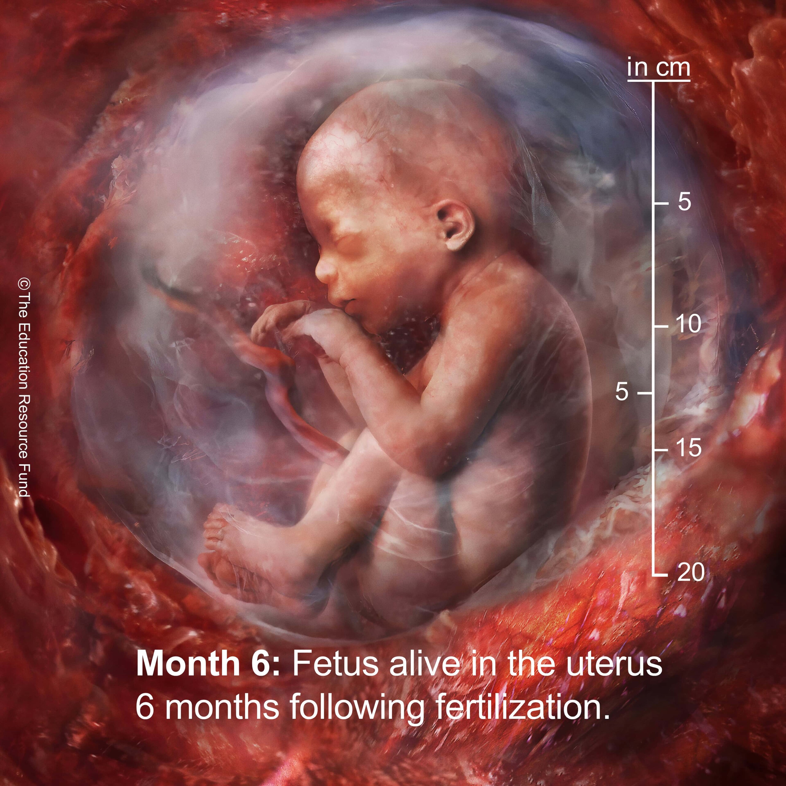
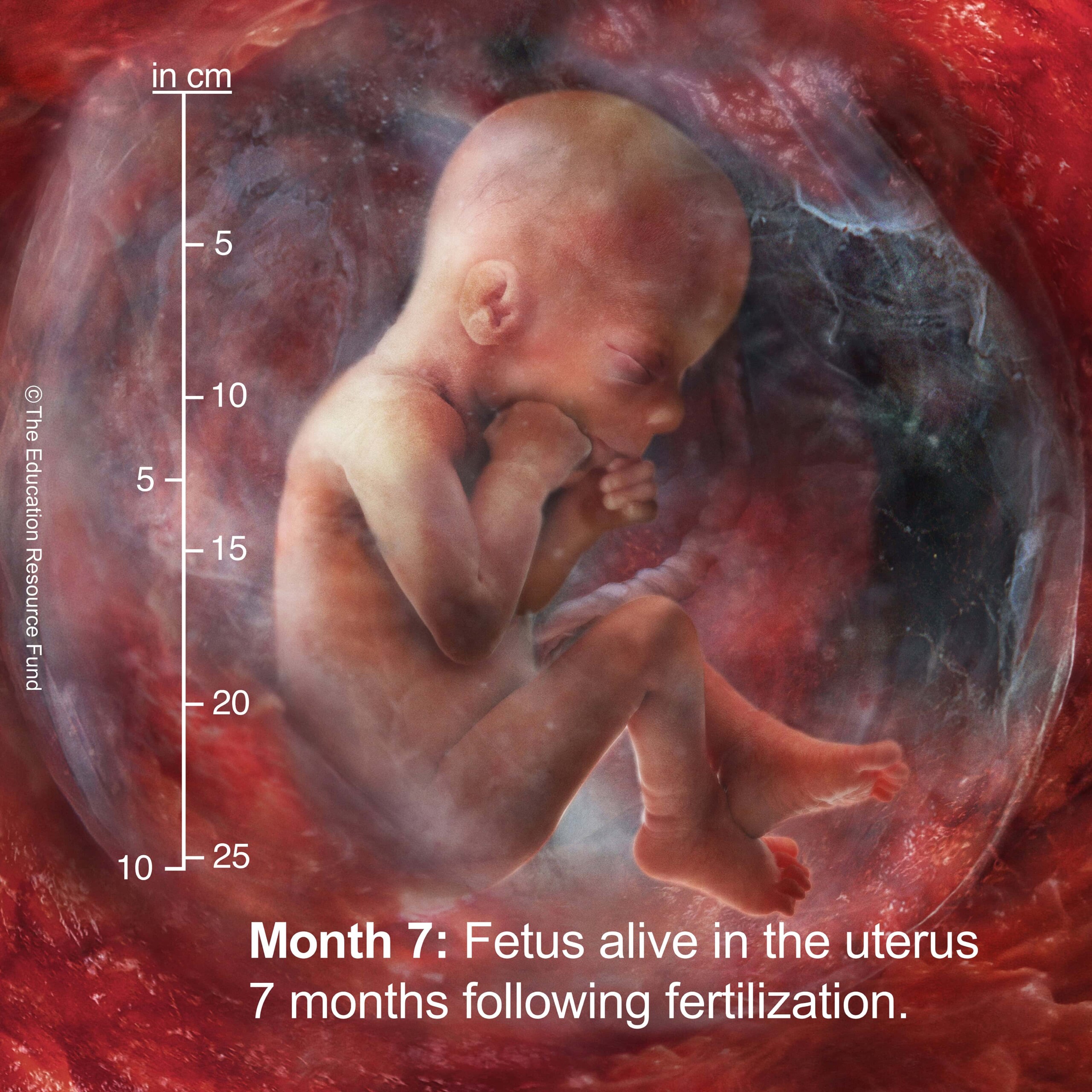
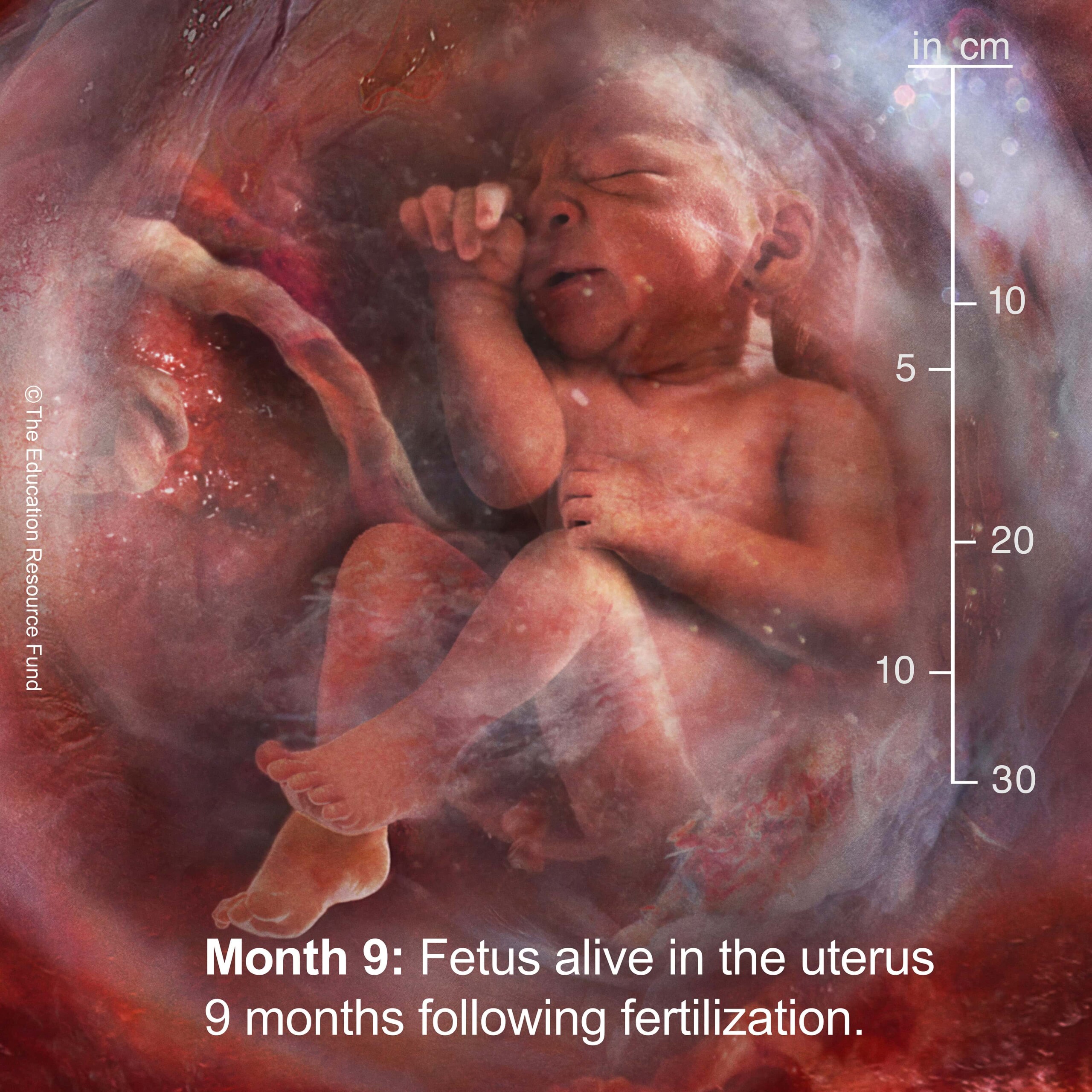
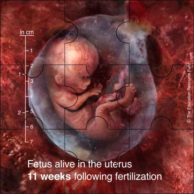
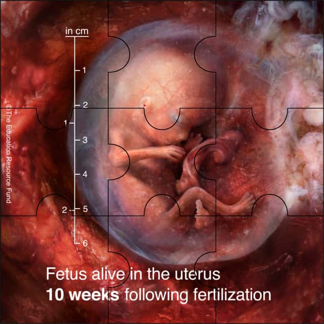
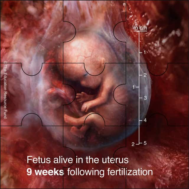
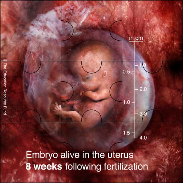
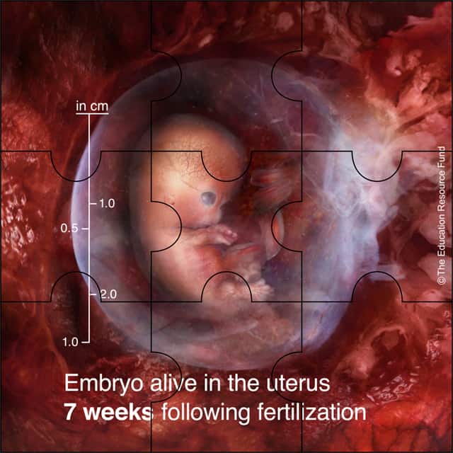
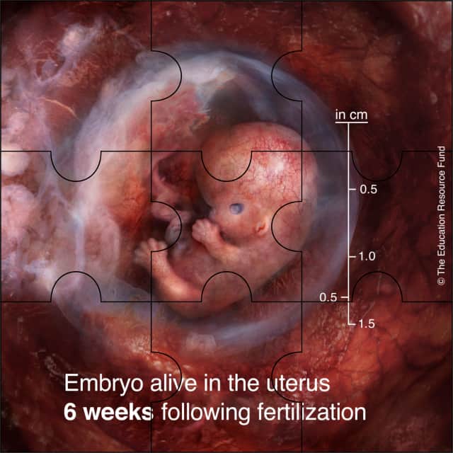
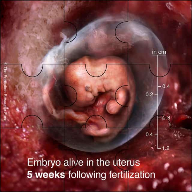
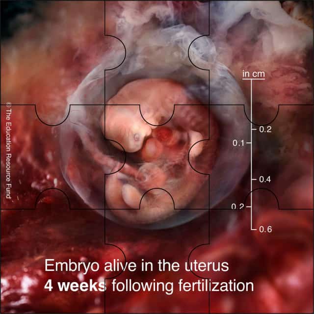
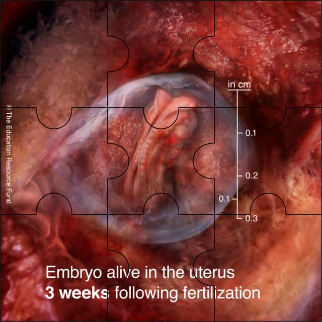
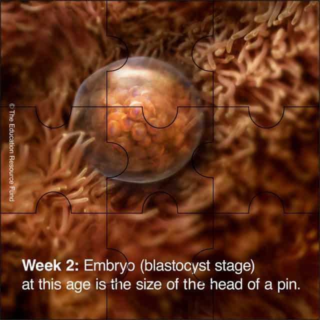
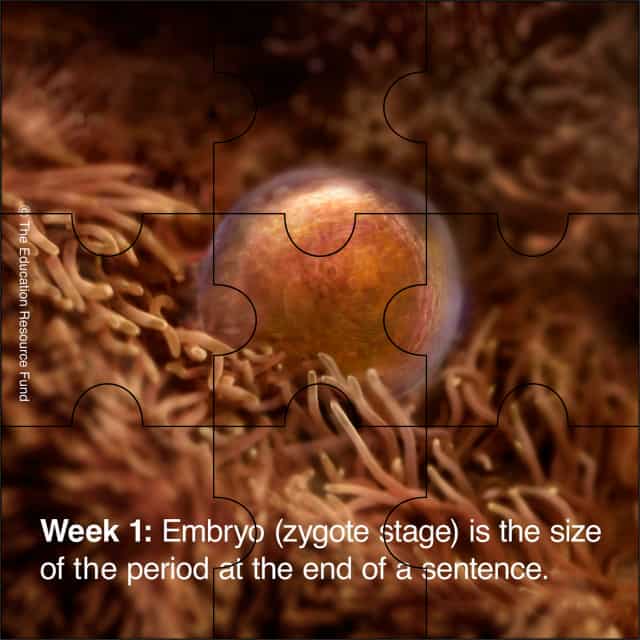
Unless otherwise noted, all embryonic and fetal ages in Education Resource Fund curricular materials are estimated in weeks/months following fertilization.
For Immediate Release
March 5 2024
The Education Resource Fund (ERF) recently announced an extraordinary new series of pregnancy-related science documentaries which illustrate the biology of prenatal development using sophisticated medical imaging technologies and procedures which enable researchers to visualize embryos and fetuses, alive in the uterus, with never-before-seen clarity. ERF is a science foundation which produces films and other curricular materials that are now available in an app ...
The Intricately Interactive Choreography of Conception
1. Male initiated: Up to 600 million sperm are deposited in the birth canal, of which only 200 reach the fertilization site in the uterine tube. The Developing Human: Clinically Oriented Embryology, 10th ed., K. Moore et al., Elsevier (2016), pp. 25-26.
2. Male initiated: An enzyme produced by...
Award-winning prenatal science website:
2024 Creative Excellence Award
Website Design
2024 dotComm Awards
Honorable Mention
2024 Horizon Interactive Awards
Bronze Winner
2025 Communicator Awards
Award of Distinction
ERF’s See Baby Grow app content has been viewed more than 18.2 million times as of July 7, 2025. More than 2,700 comments have been posted. Viewers are from at least 173 countries.

Award-winning prenatal science mobile app:
© See Baby Grow App
To obtain the See Baby Grow App for both Apple (iOS) and Google Play (Android), download from the Apple App Store at the foregoing QR Code, or this link:


2024 Horizon Interactive Awards
Gold Winner

2024 Webby Awards
Honoree
Apps and Software
2025 Communicator Awards
Award of Distinction
2023 Best Mobile App Awards
Nominee

Embryoscopy, fetoscopy, and high-resolution ultrasound imagery, showing embryonic and fetal development
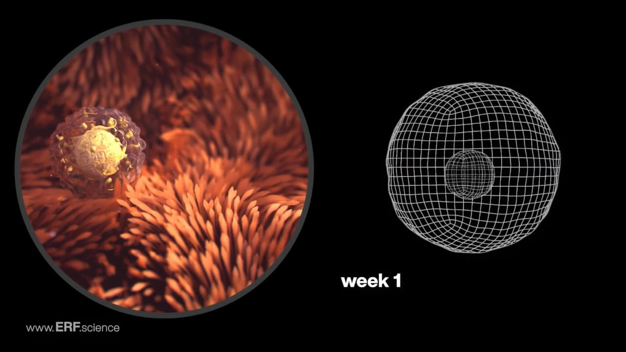
Week 1 – Embryonic & Fetal Video Clips
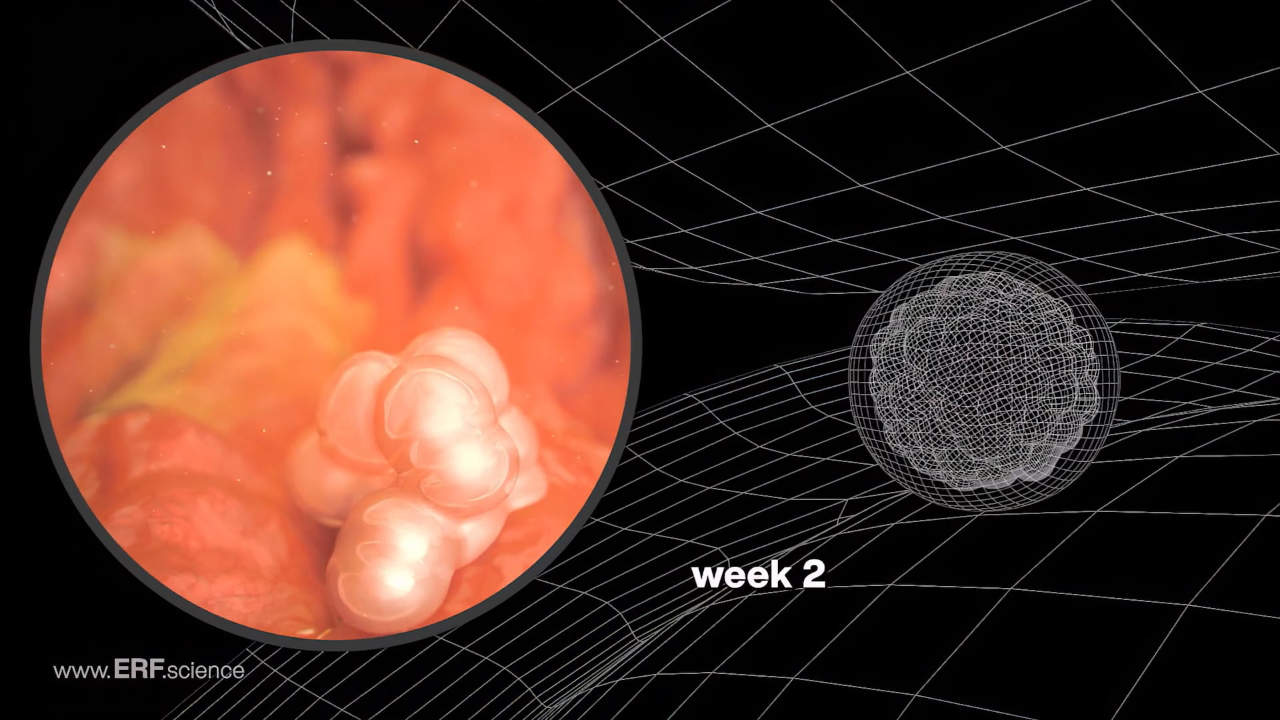
Week 2 – Embryonic & Fetal Video Clips
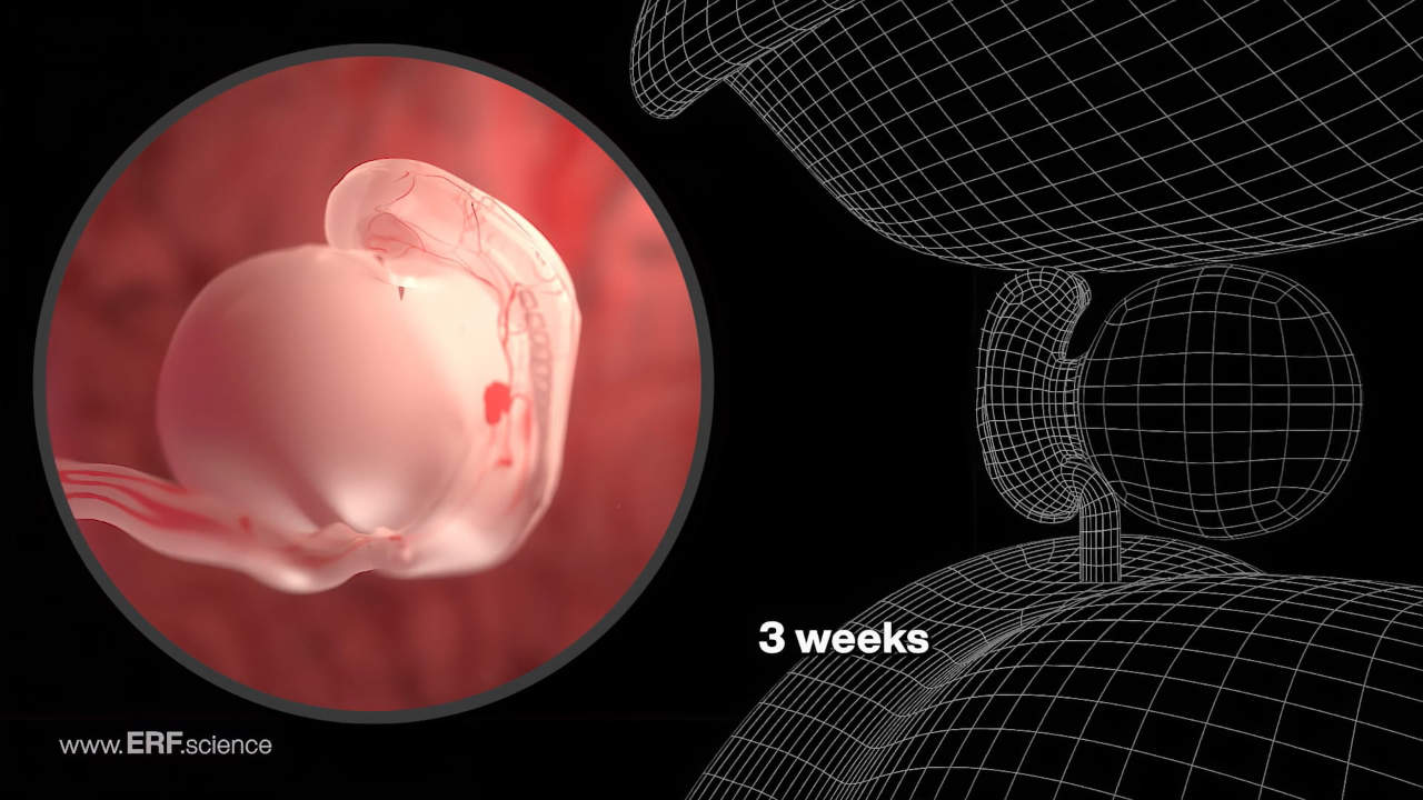
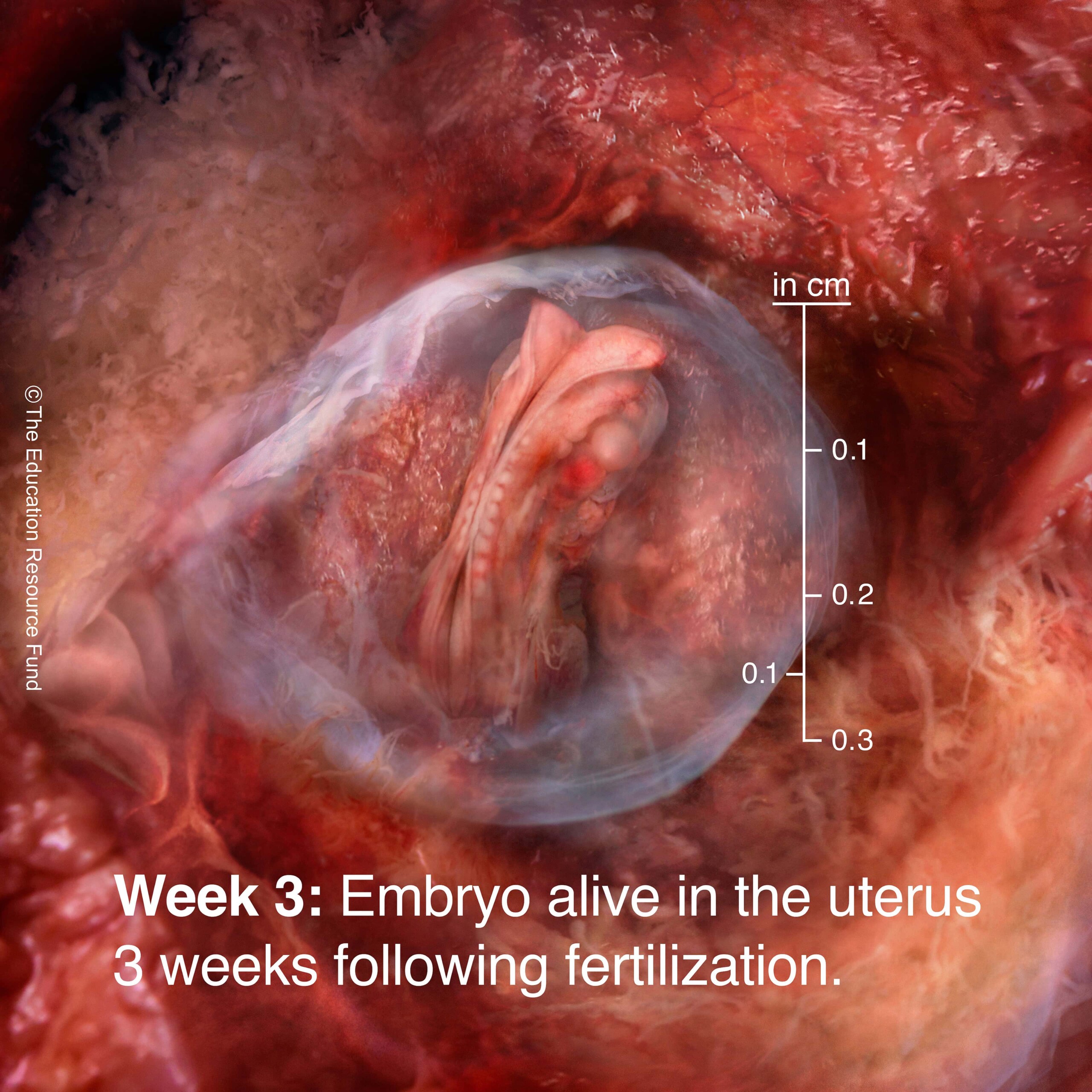
Week 3 – Embryonic & Fetal Video Clips
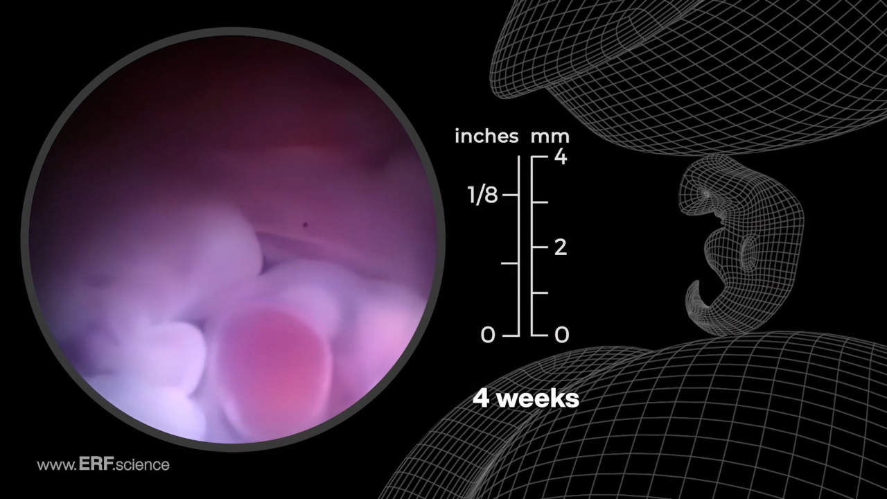
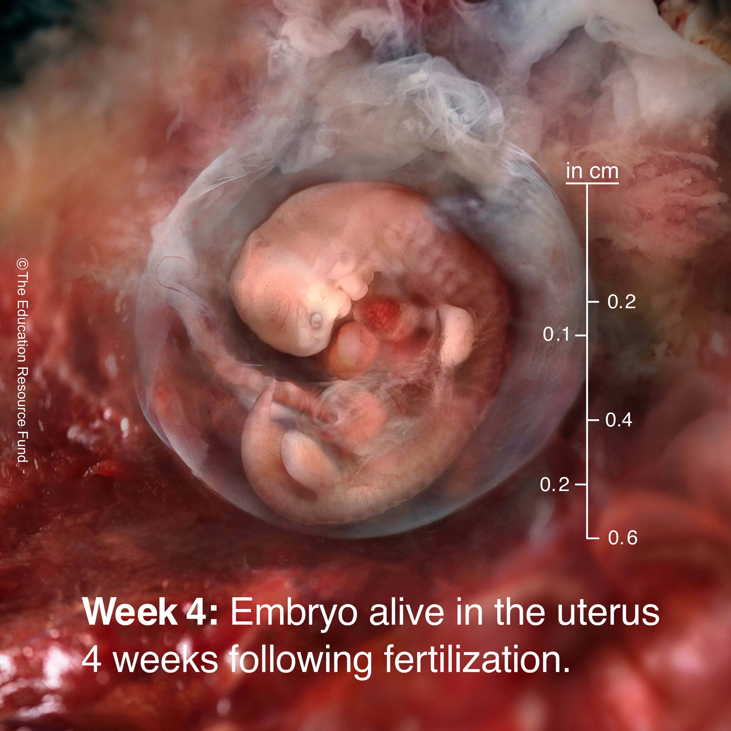
Week 4 – Embryonic & Fetal Video Clips
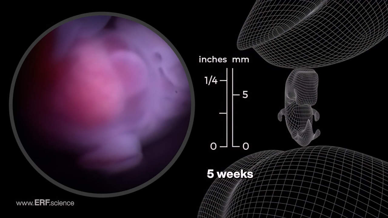
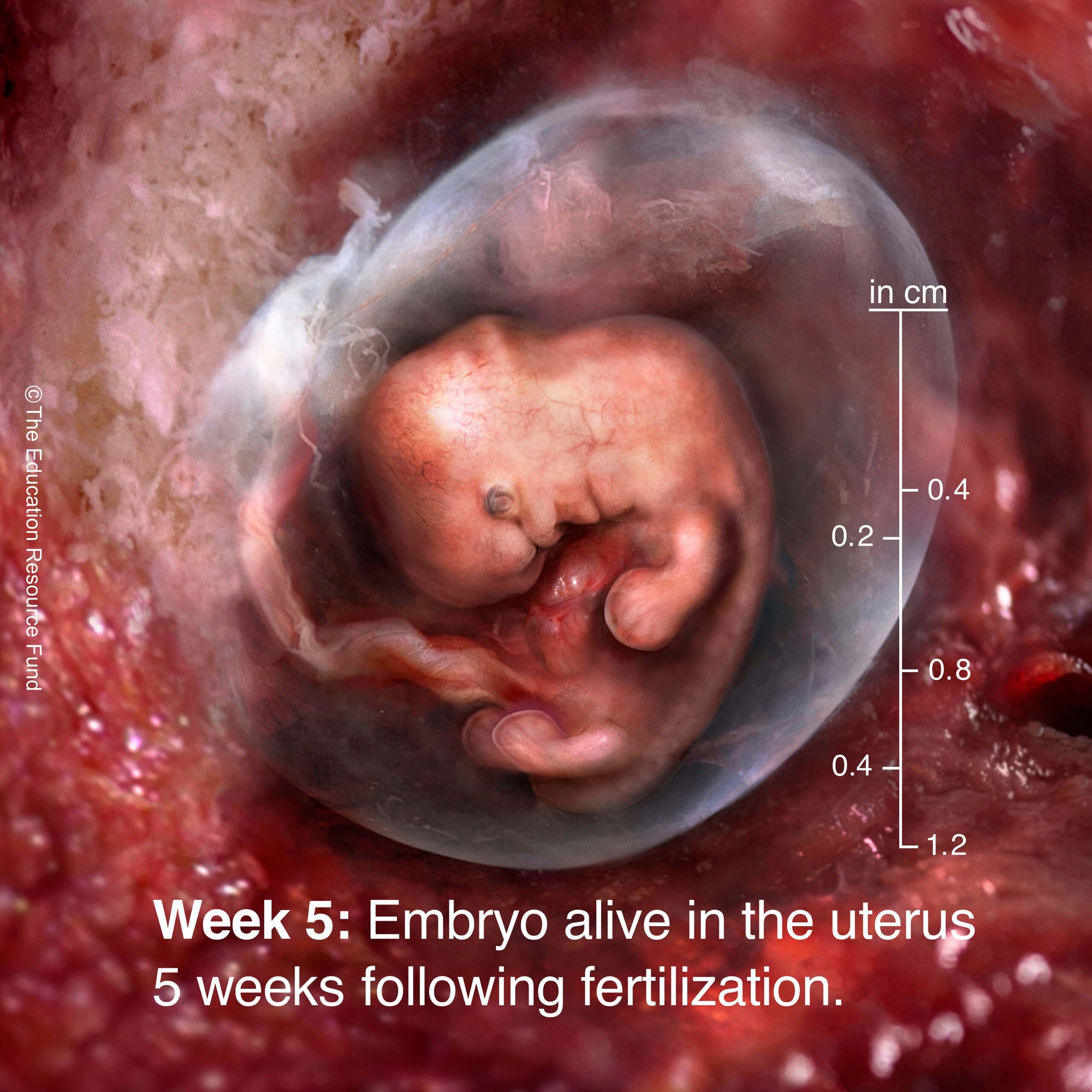
Week 5 – Embryonic & Fetal Video Clips
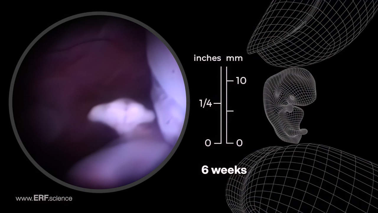
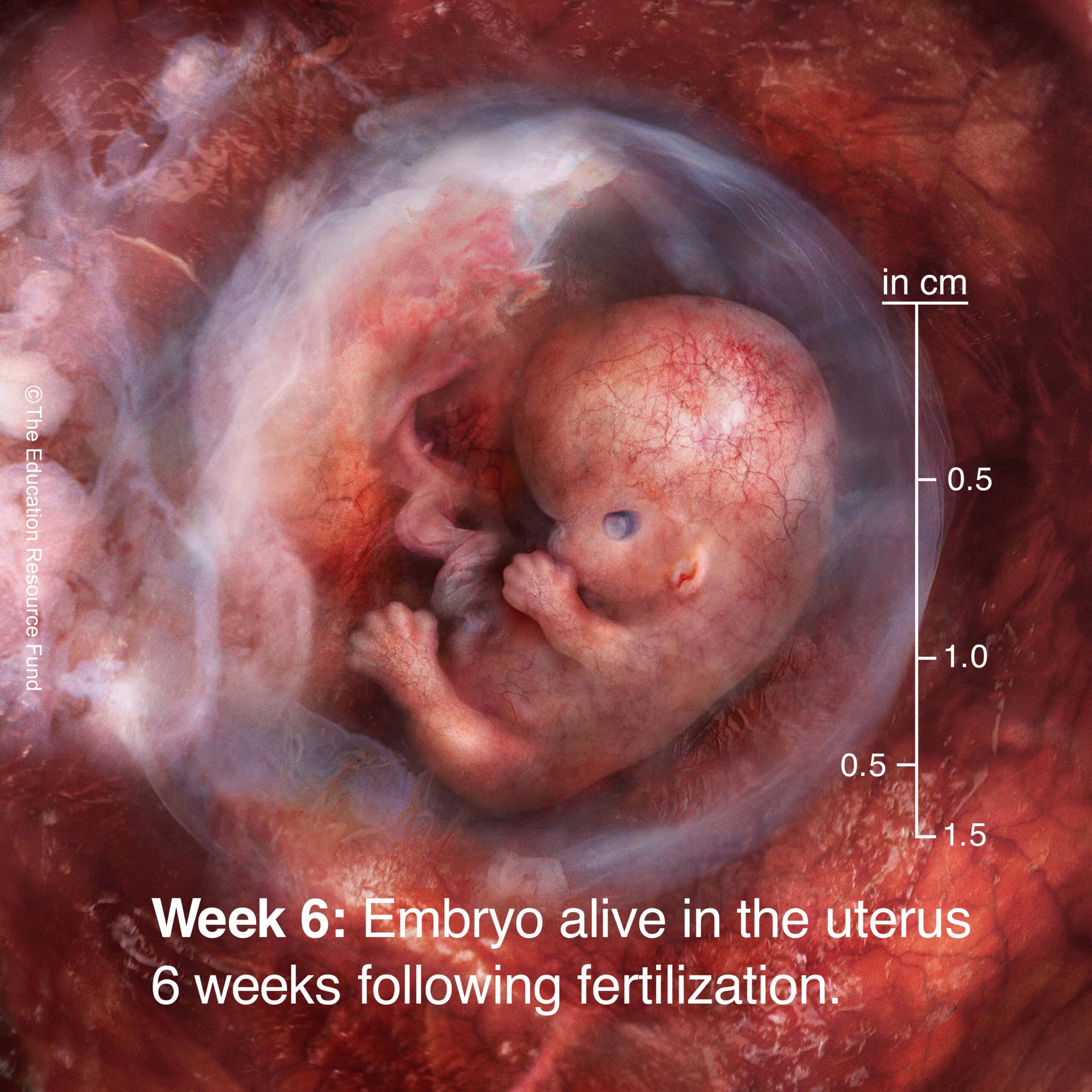
Week 6 – Embryonic & Fetal Video Clips
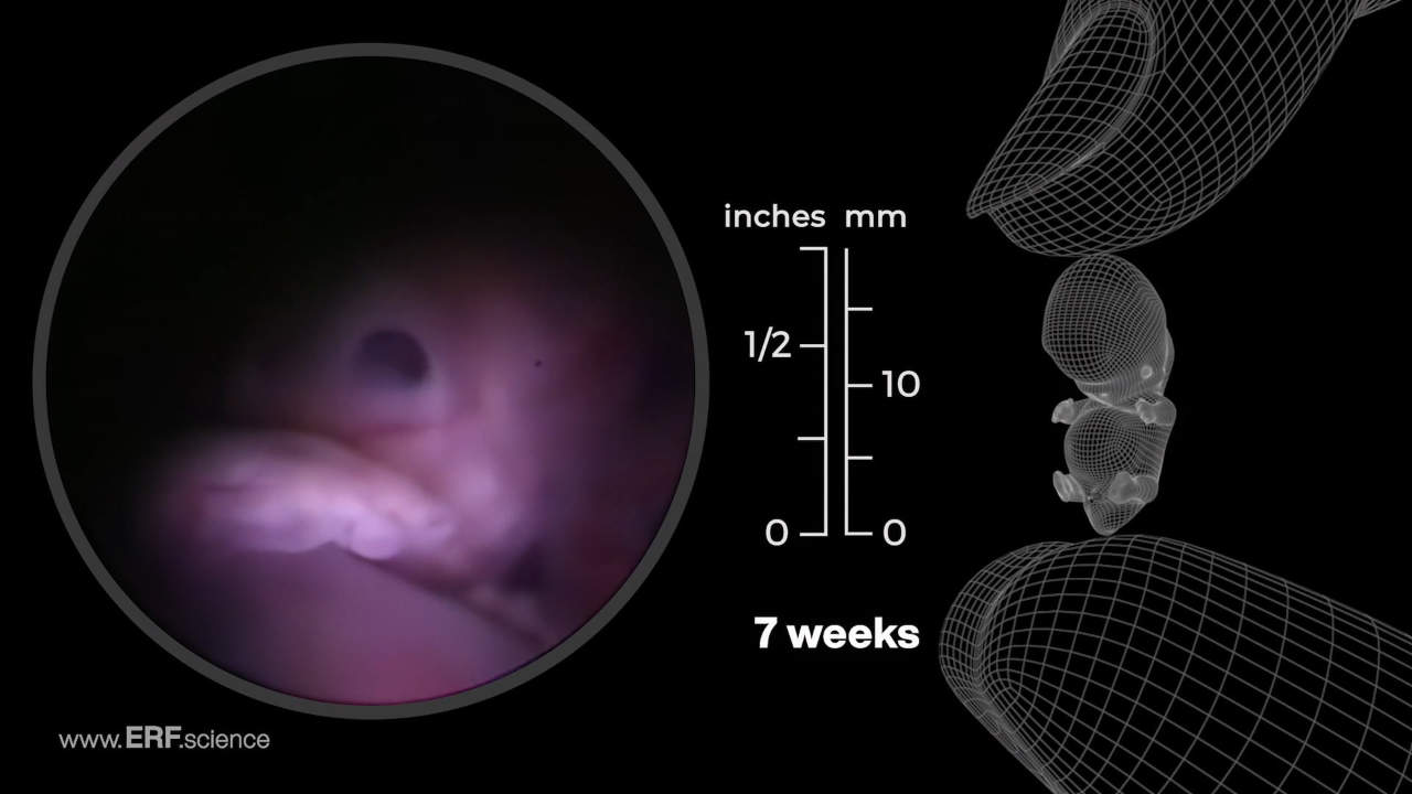
Week 7 – Embryonic & Fetal Video Clips
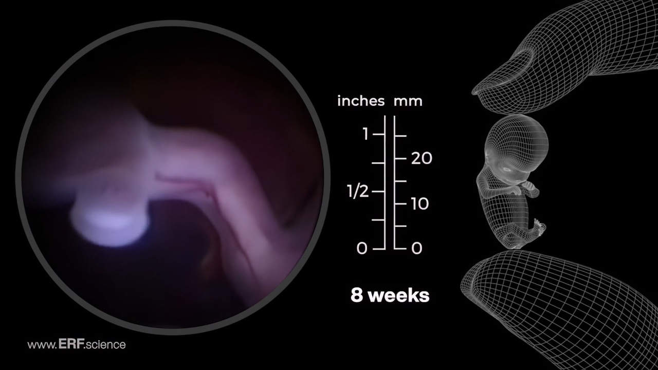
Week 8 – Embryonic & Fetal Video Clips
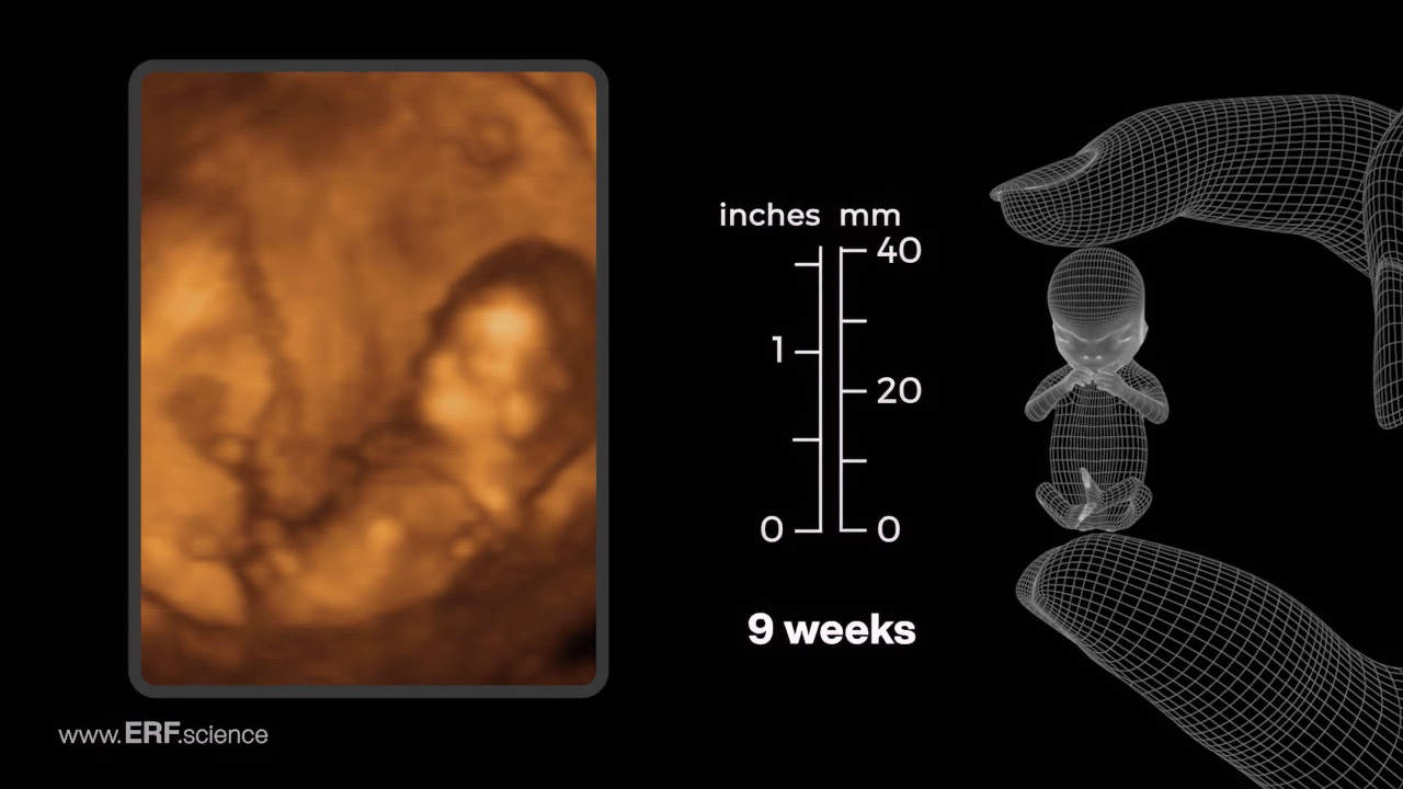
Week 9 – Embryonic & Fetal Video Clips
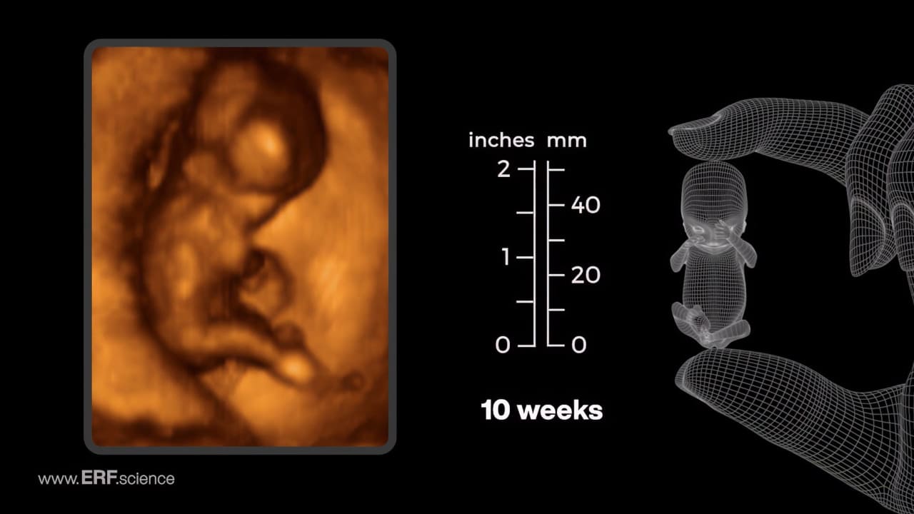
Week 10 – Embryonic & Fetal Video Clips
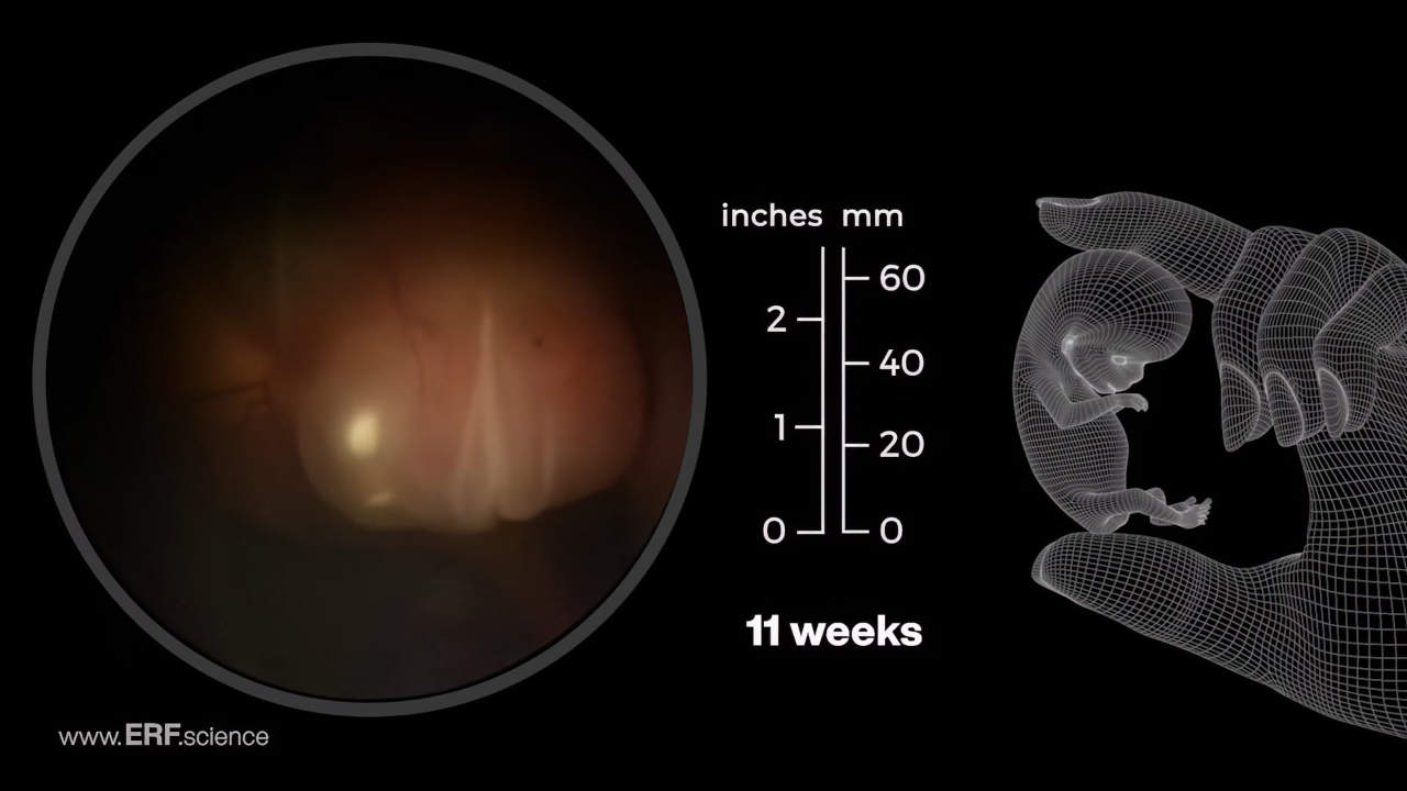
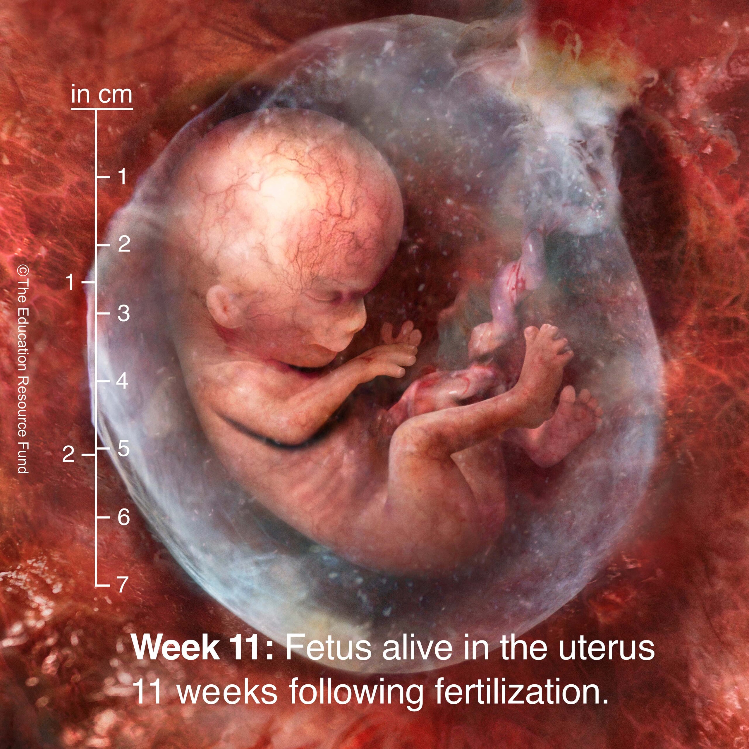
Week 11 – Embryonic & Fetal Video Clips
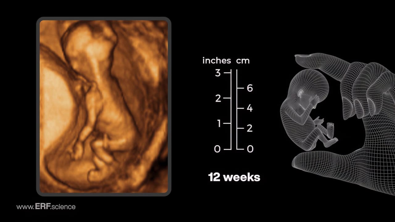
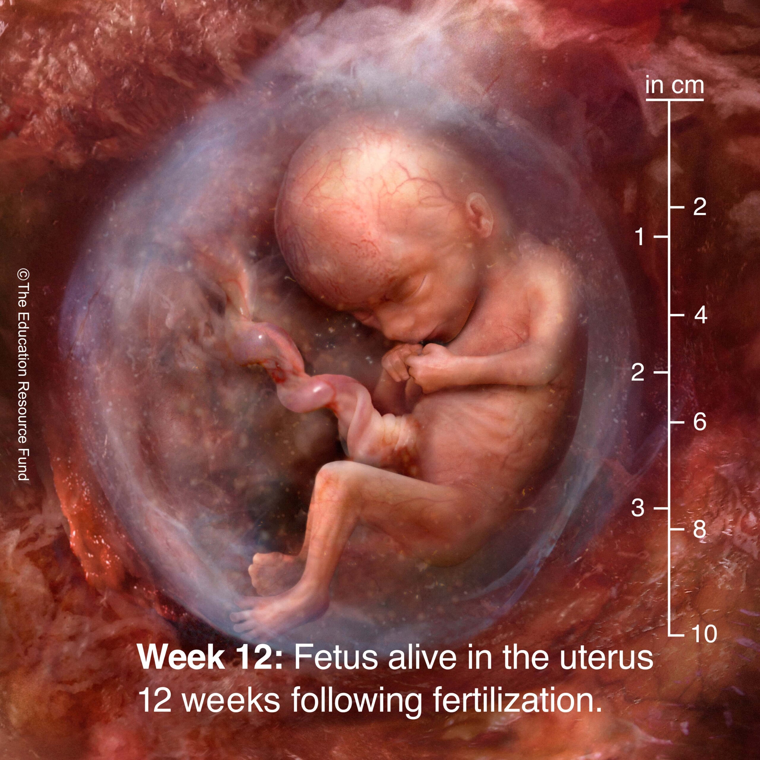
Week 12 – Embryonic & Fetal Video Clips
Subtitles in 92 languages for ERF video "The Science of Life Before Birth"
Choose Your Preferred Subtitle Language Here:
We have prepared the script of “The Science of Life Before Birth” in 92 different languages. You can download a PDF of each of those translations by following the links below.


See Baby Grow app video
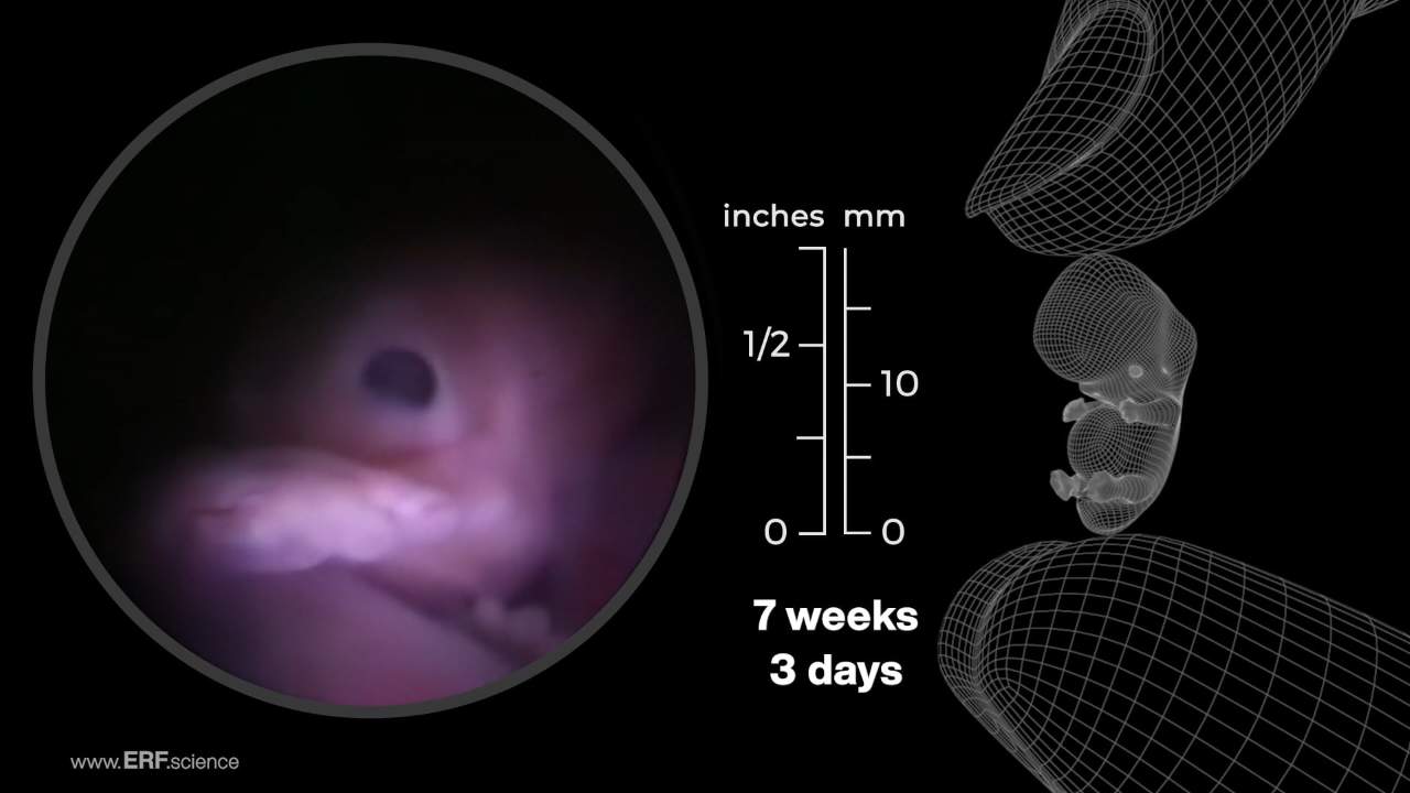
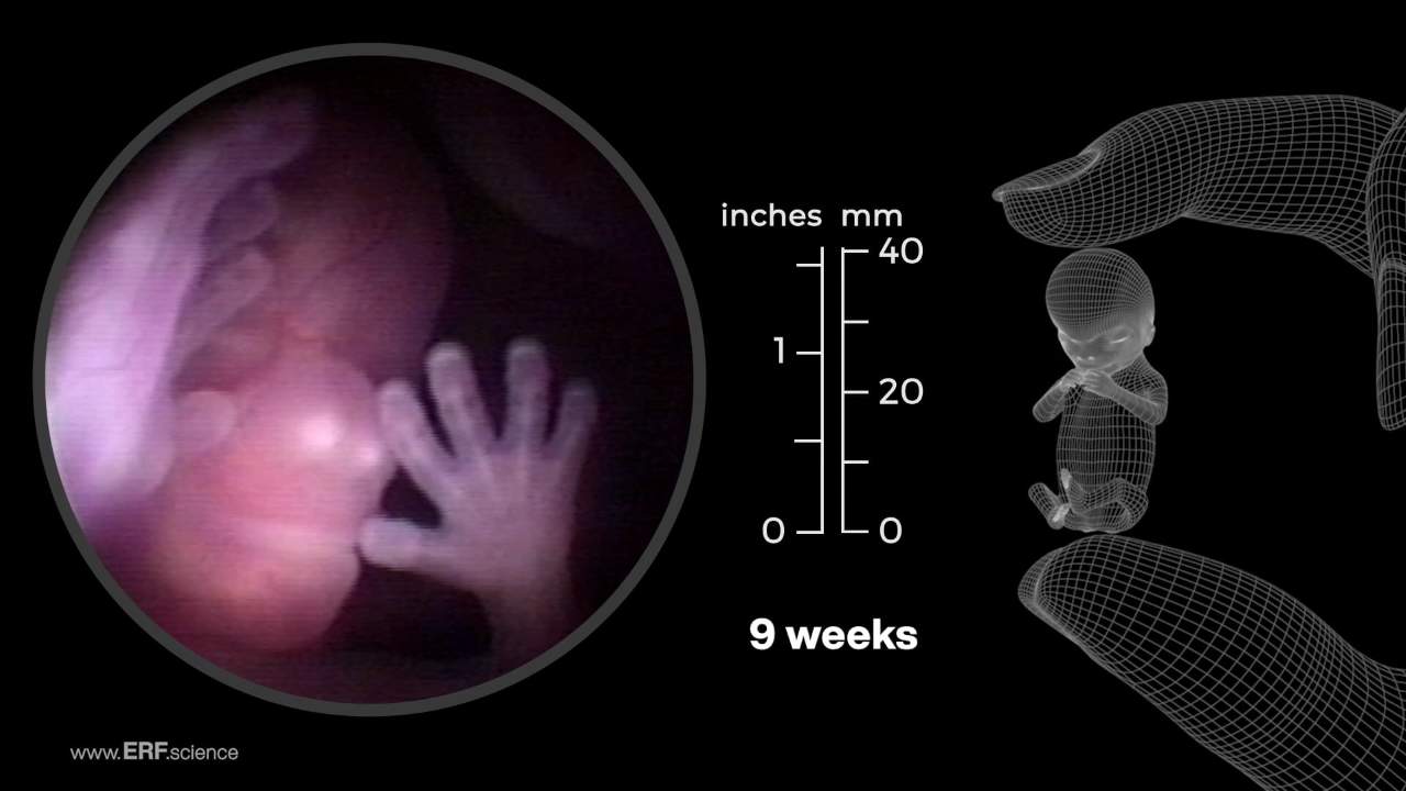
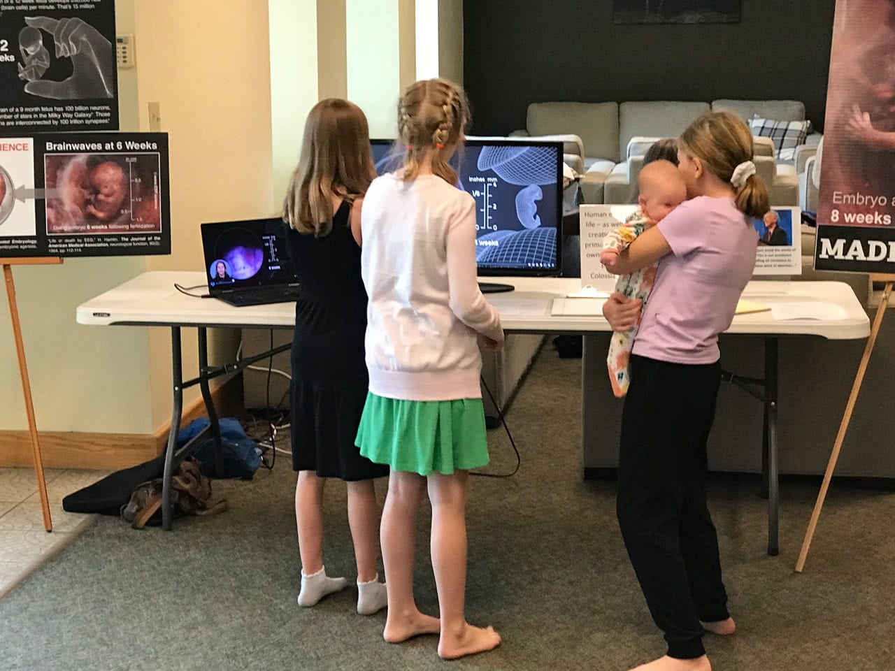
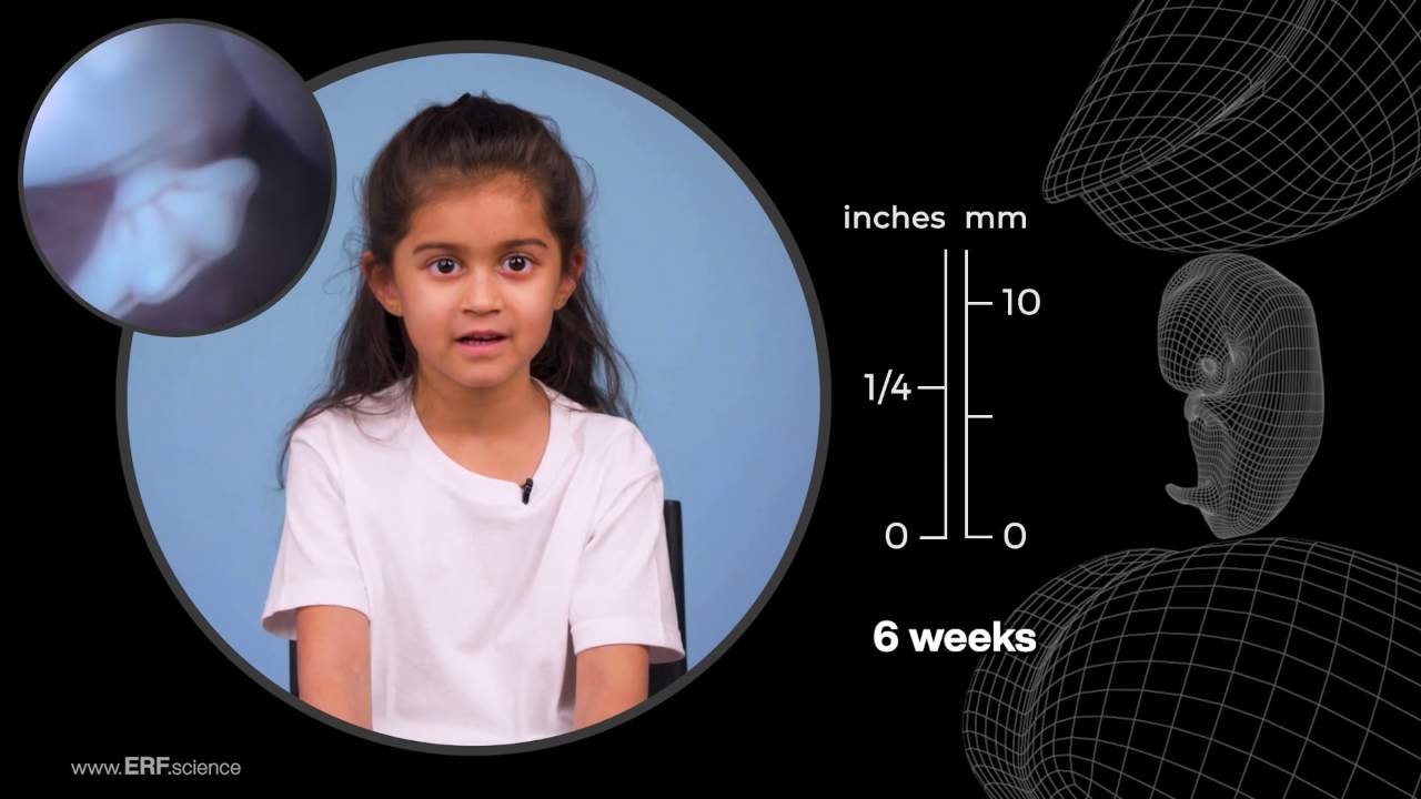
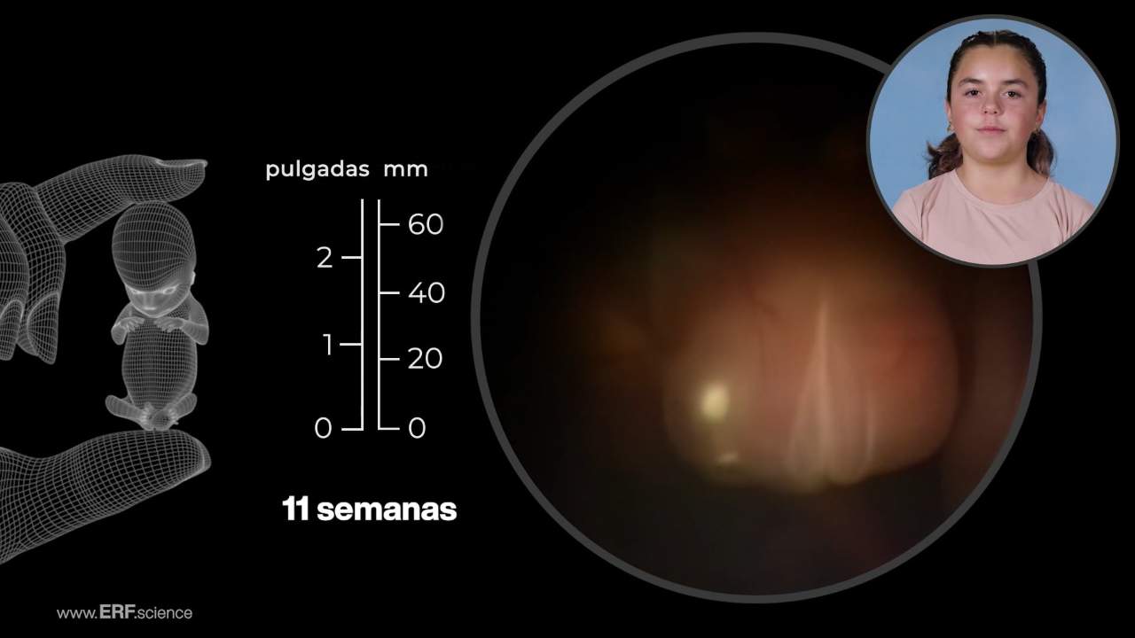
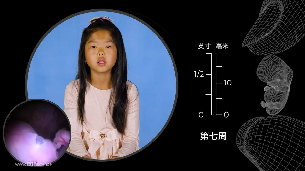
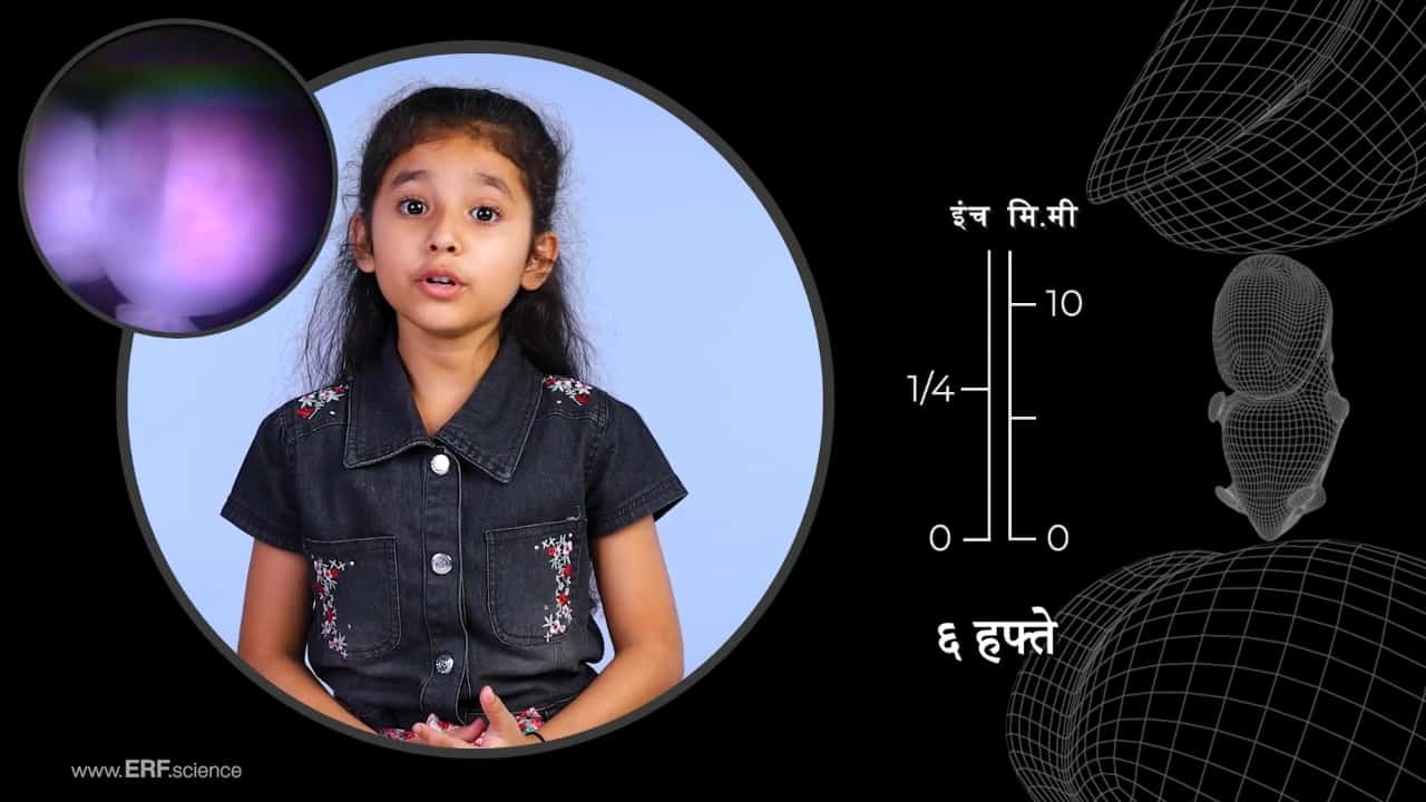
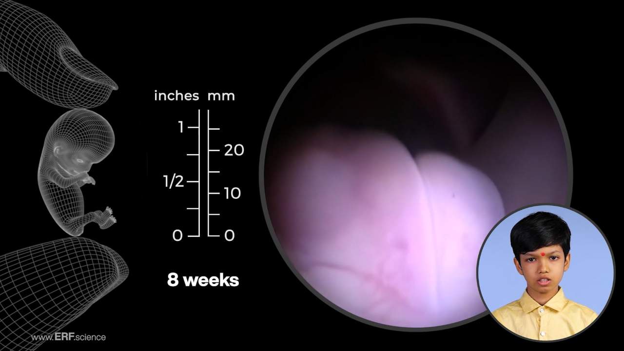
The See Baby Grow app video depicts embryos and fetuses, alive in the uterus, throughout every stage of pregnancy. These preborn babies have been scanned using embryoscopy and fetoscopy medical imaging technology, as well as high-resolution, research-grade sonography. The narration describes developmental anatomy and physiology as it unfolds through all three trimesters of pregnancy.
Post the following prenatal development facts on your social media:

The ERF prenatal videos featured at www.ERF.science are updated and expanded variants of the science documentary titled "The Biology of Prenatal Development."
Biology of Prenatal Development Film Awards

CINE Golden Eagle

Platinum Remi Award

Grand Remi – “Best of Show”

Grand Remi – “Best of Show”

Grand Remi

Award of Excellence

Silver Telly Award
“The developmental period before birth is increasingly understood as a time of preparation during which the developing human acquires the many structures, and practices the many skills, needed for survival after birth. As our understanding of early human development advances, so too will our ability to enhance health––both before and after birth.”
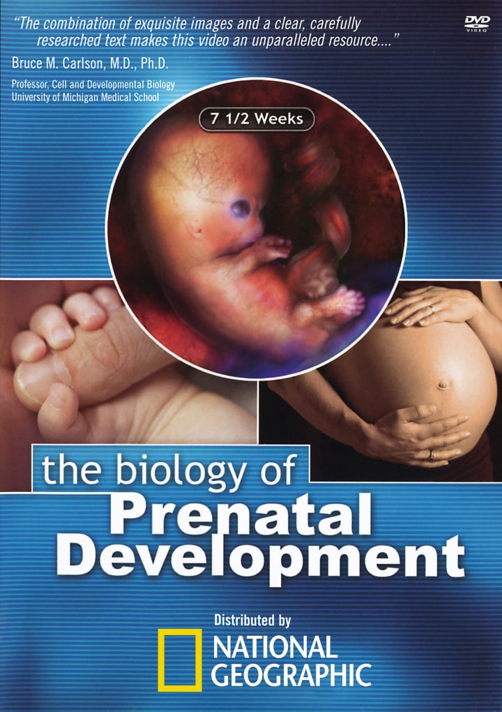
The Biology of Prenatal Development, a documentary film originally distributed by the National Geographic Society
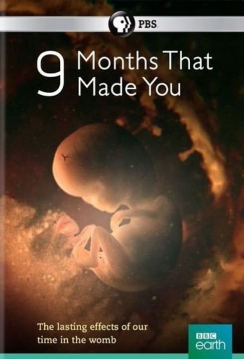
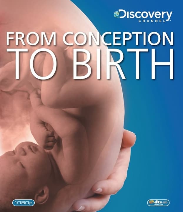
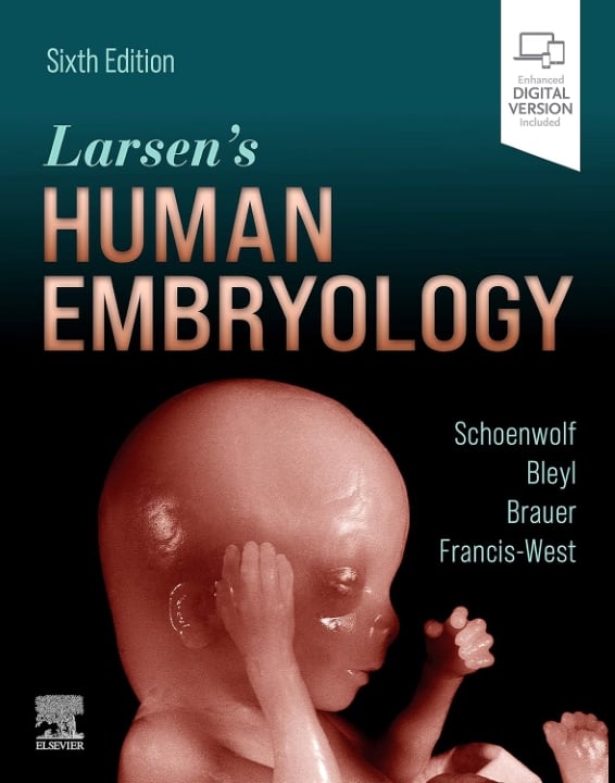
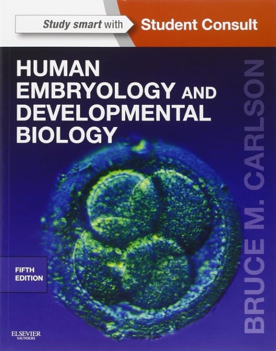
The following science documentaries, medical textbooks, and medical journal articles are among the many educational resources which provide useful information regarding the biology of prenatal development:
Recommended Science Documentaries


Recommended Medical Textbooks


Medical Journal Articles
IMAGING THE UNIMAGINABLE
The Education Resource Fund’s (ERF) composited embryonic and fetal pictures are derived from many smaller images, “stitched” together in much the same manner employed by NASA to combine satellite photo “tiles,” in a process which forms a large “mosaiced” image. Many research institutions use related technology to image otherwise unimageable (and unimaginable) objects and processes.
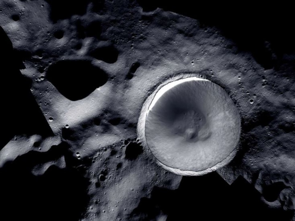 A mosaic image of the lunar South Pole, with individual photo tiles electronically stitched together to create one enormous panoramic view. Photo credit NASA
Note: ERF does not use animated imagery to depict any structures or processes which are capable of being filmed, photographed, or scanned.
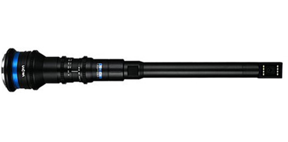
ENDOSCOPES
ERF’s embryo and fetus imagery was initially derived by teams of physician researchers and clinicians employing endoscopy (and its subsets, embryoscopy and fetoscopy) to diagnose and treat prenatal disorders in utero.
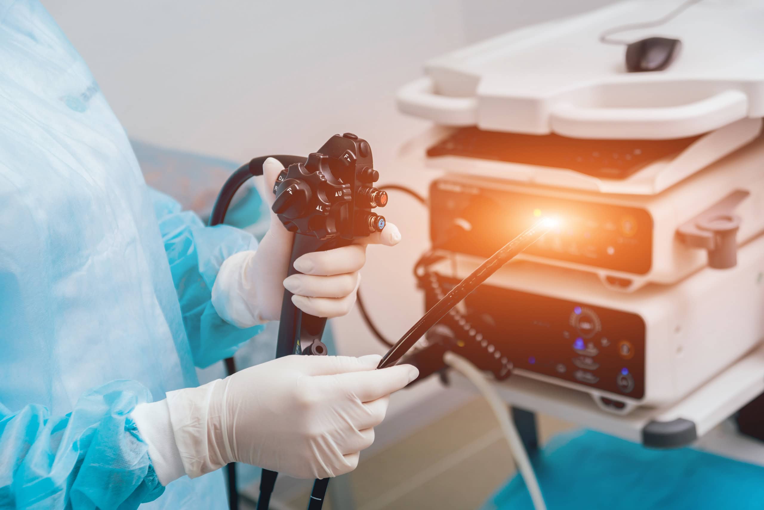 An endoscope with related equipment
Endoscopes are medical imaging devices which permit high-resolution observation of tissues and processes inside the human body. Prenatally, they can be used to produce minimally invasive scans imaged through, but from outside, the amnion. When clinically necessary, more invasive scans may be performed by surgically entering the abdominal cavity, uterus and amniotic sac. At the distal end of these instruments is an objective lens designed for imaging. At the proximal end is an eyepiece, or sensor, which enables viewing.
HOW THEY WORK
These scopes generally consist of a tube which encloses a relay lens system (in rigid endoscopes) or a fiber bundle (for fiber-optic, or flexible, endoscopes) for illumination and to transmit an image from the objective lens inside the body to the proximal end outside.
Said differently, endoscopes use optical elements to direct light to the area sought to be illuminated and transmit the resulting image to the eye or detector. Rigid endoscopes generally offer superior resolution or magnification. But an endoscope’s objective lens is only approximately 1/5 of an inch in diameter, and this relatively small size substantially narrows the observer’s field of view (even with the addition of supplemental lenses such as “negative” or “prism” optics, etc.).
CONSTRAINTS
This limitation is further compounded by the need to use the scope in very confined spaces, with only short distances separating the objective lens from the anatomical structures being imaged. Consequently, only a small segment of the embryo or fetus is observable at any point along the timeline of the scan.
An endoscope’s construction must also accommodate frequently conflicting design considerations. The resulting compromises can involve not only fields of view, but depths of field (meaning thickness of the plane of focus) and image illumination and magnification, as well as distortion issues (i.e., stretched or compressed perspective), etc.
WORK-AROUNDS
Therefore, to produce a high-quality, single image of the entire embryo or fetus, large numbers of smaller, more detailed pictures must be joined together in a manner suggestive of the process by which puzzle pieces are assembled to form a completed picture.
This technique employs a complex proprietary process which combines segmental scans to create a final, multi-source composited image. The resulting picture is digitally adjusted to preserve each segment’s original color, resolution, contrast, proportions, illumination, etc. Technicians also correct for vignetting (image degradation involving content loss at the periphery of the frame).
MAGNETIC RESONANCE IMAGING & ULTRASOUND
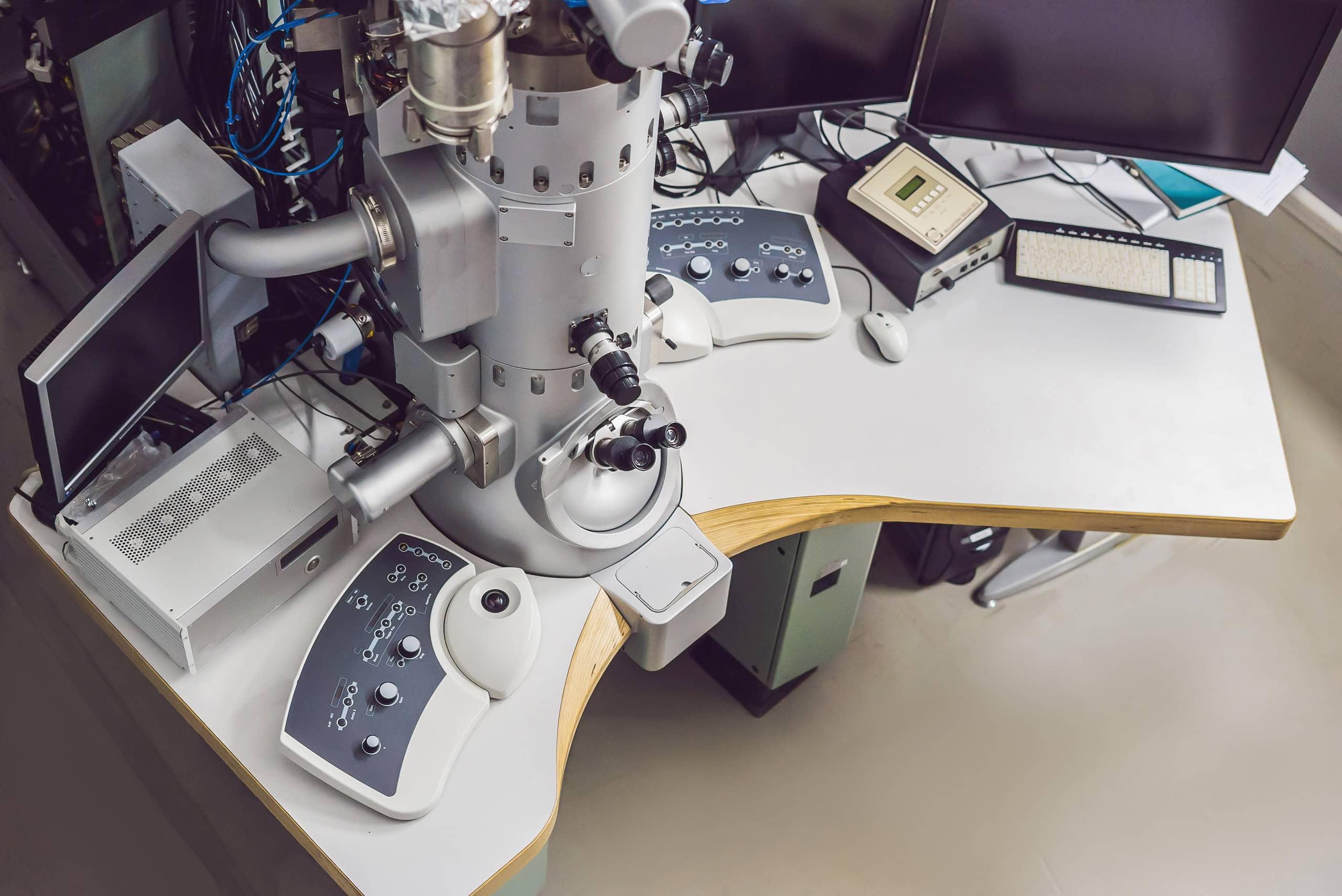
The British medical journal Lancet has published a prenatal magnetic resonance imaging (MRI) study similarly involving the creation of 3D pictures to diagnose and treat congenital heart problems in utero. The BBC reports that “A series of 2D pictures of the heart are taken from different angles using an MRI machine” to image the fetus.
 Magnetic Resonance Imaging (MRI) equipment with digital images of scanned tissue.
The story explains that “Sophisticated computer software pieces the images together, adjusts for the beating of the heart and builds … [a] 3D image of the heart.” A pediatric cardiologist describes the resulting 3D images as “beautiful.”
This MRI research is part of a fetal diagnostic project which is also exploring scans using “four ultrasound probes at the same time – current scans use one – to get a more detailed picture.” This process produces a more wholistic composited image.
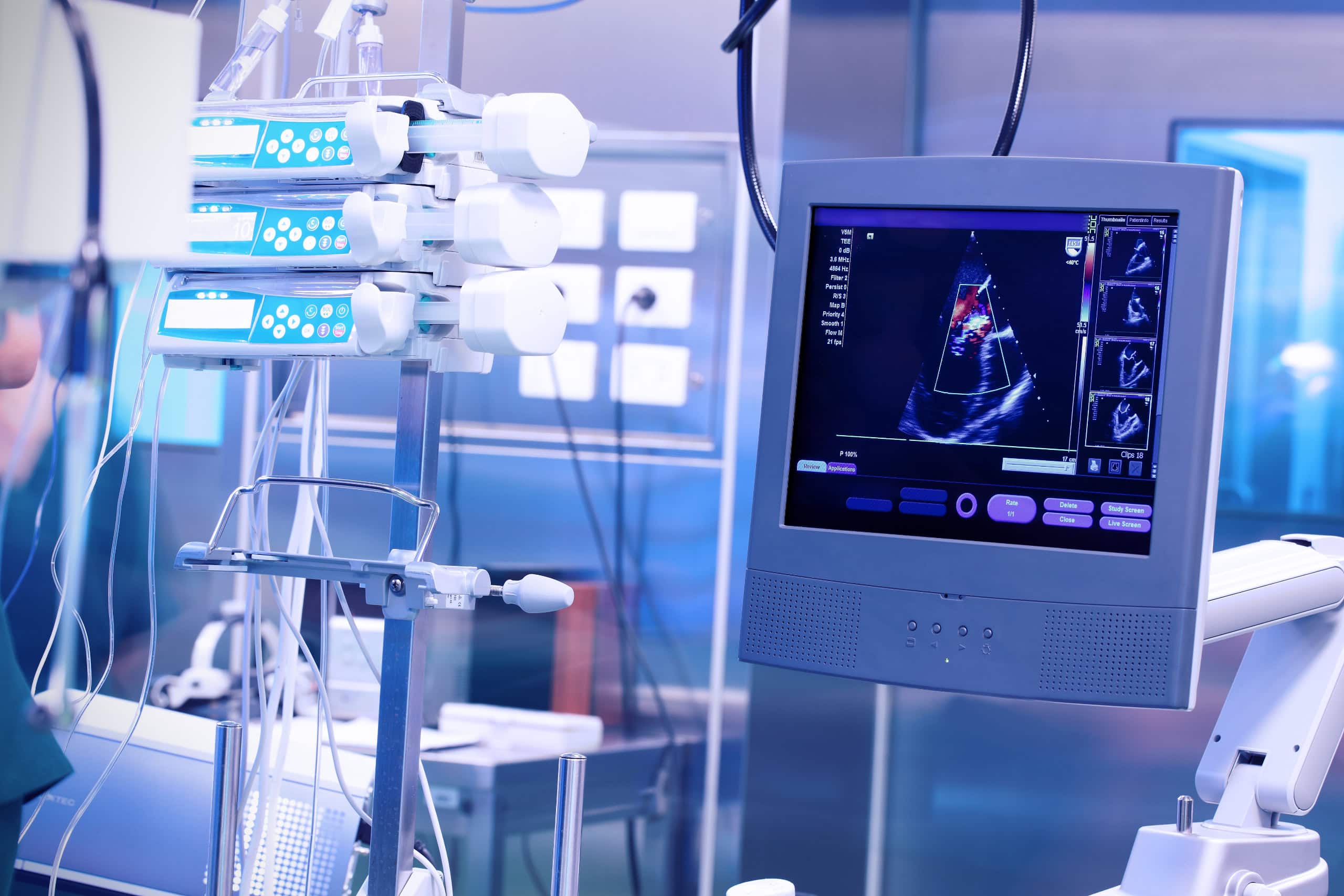 Research-grade ultrasound scanning equipment.
NASA COMPOSITES IMAGERY (SINGLE MEDIUM)
ERF’s imaging process is conceptually similar to the technologies used by the National Atmospheric and Space Administration (NASA) to produce wide-area satellite images of the earth’s surface. Until the launch of the Deep Space Climate Observatory Satellite (DSOVR), which now orbits the earth at a distance of one million miles, NASA had no camera positioned sufficiently far from our planet to capture the globe’s entire sunlit surface in a single photograph.
As previously noted, an endoscope’s objective lens must also operate too near to an embryo or fetus to permit its entire anatomy to be imaged in a single frame. This same constraint complicates the capture of satellite imagery. Previous pictures of the earth could, therefore, only be created using digital “stitching” technology to make one large composite image from many smaller segments. Scientists sometimes describe this final image (or “data set”) as a “mosaic,” comprised of large numbers of individual tiles.
HYBRID IMAGERY (MULTI-MEDIA)
A satellite picture can also be augmented by aerial photography (cameras on aircraft operating within the earth’s atmosphere) to improve resolution. Hybrid images of this sort can be created by superimposing black and white imagery (for still higher resolution) over color pictures of the same area, the latter to optimize chromic (color) fidelity.
In this connection, the scientific press reports that the Landsat Image Mosaic of Antarctica (LIMA) “combined over one thousand precise, calibrated satellite images with other data from the continent’s surface to create a single picture of the entire continent.” The high magnification factor (think telephoto lenses which enlarge image objects) of each of these puzzle pieces yielded a composite picture depicting more detail than could have been captured in a single photo shot with a wide-angle lens.
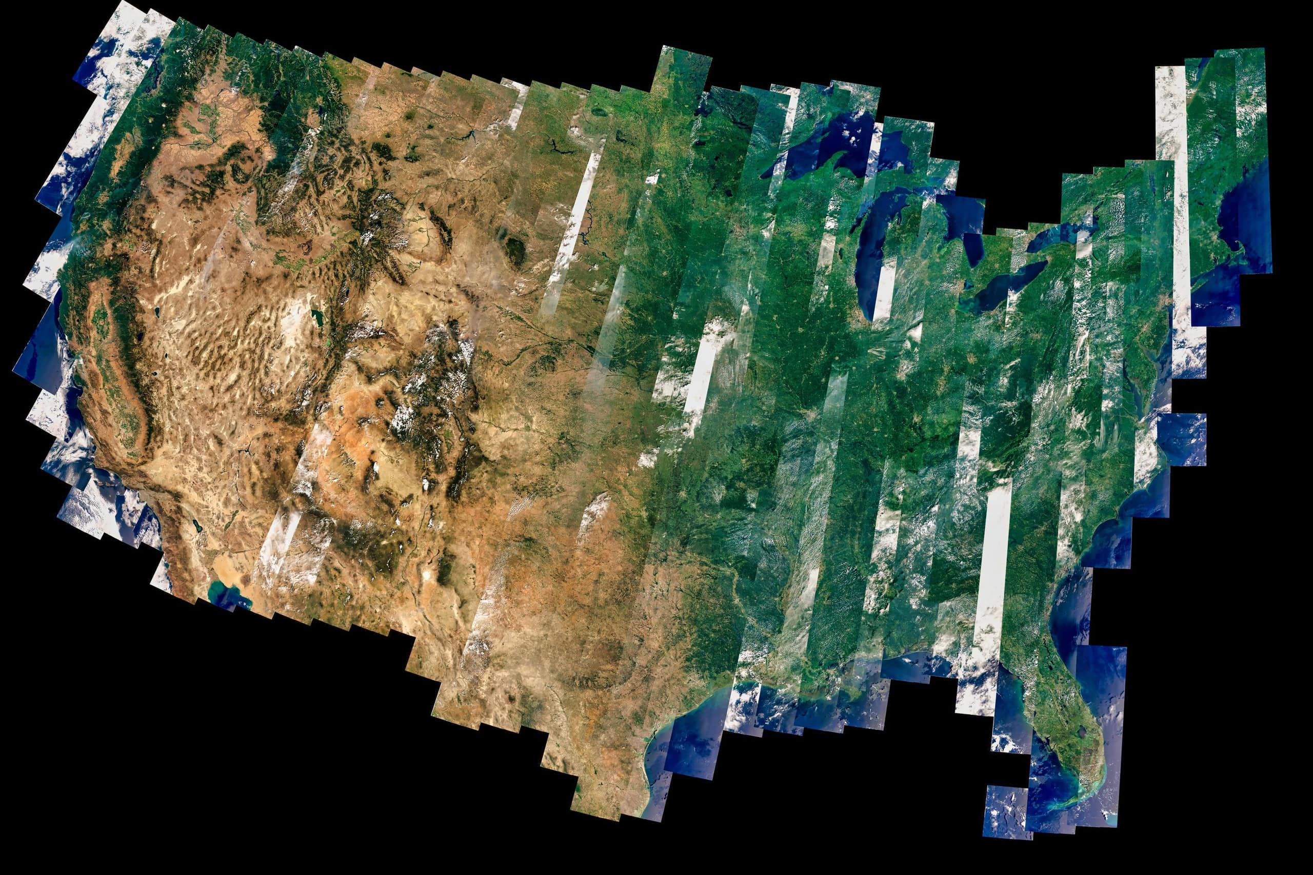 Landsat satellite cameras generate composited images of the earth’s surface during multiple passes with continuous scans per pass.
APPLICATIONS IN ASTRONOMY
NASA uses this mosaicking process to image celestial bodies of nearly every description. The Juno spacecraft made composite images of Jupiter; InSight of Mars; Cassini of Saturn; and Hubble of the Sombrero Galaxy.
An exquisitely detailed depiction of a challenging subject, whether prohibitively small or large, near or far, may involve vastly more complexity than meets the eye, and there exist nearly countless examples of comparably creative combinations of imaging techniques.
ENTAMOLOGICAL USES
NASA is not alone in its use of this imaging technique. A Wall Street Journal story, July 30, 2022, titled “Breathtaking Bugs at the Museum,” describes an exhibition of “macrophotography prints [which] makes us appreciate these small creatures’ beauty on a large scale.” The article explains the creation of large format prints “in an exhibit called ‘Extinct and Endangered, Insects in Peril.’” The imaging process is called “macrophotography,” and it took an average of “three weeks and as many as 10,000 files for the photographer to make an individual image.”
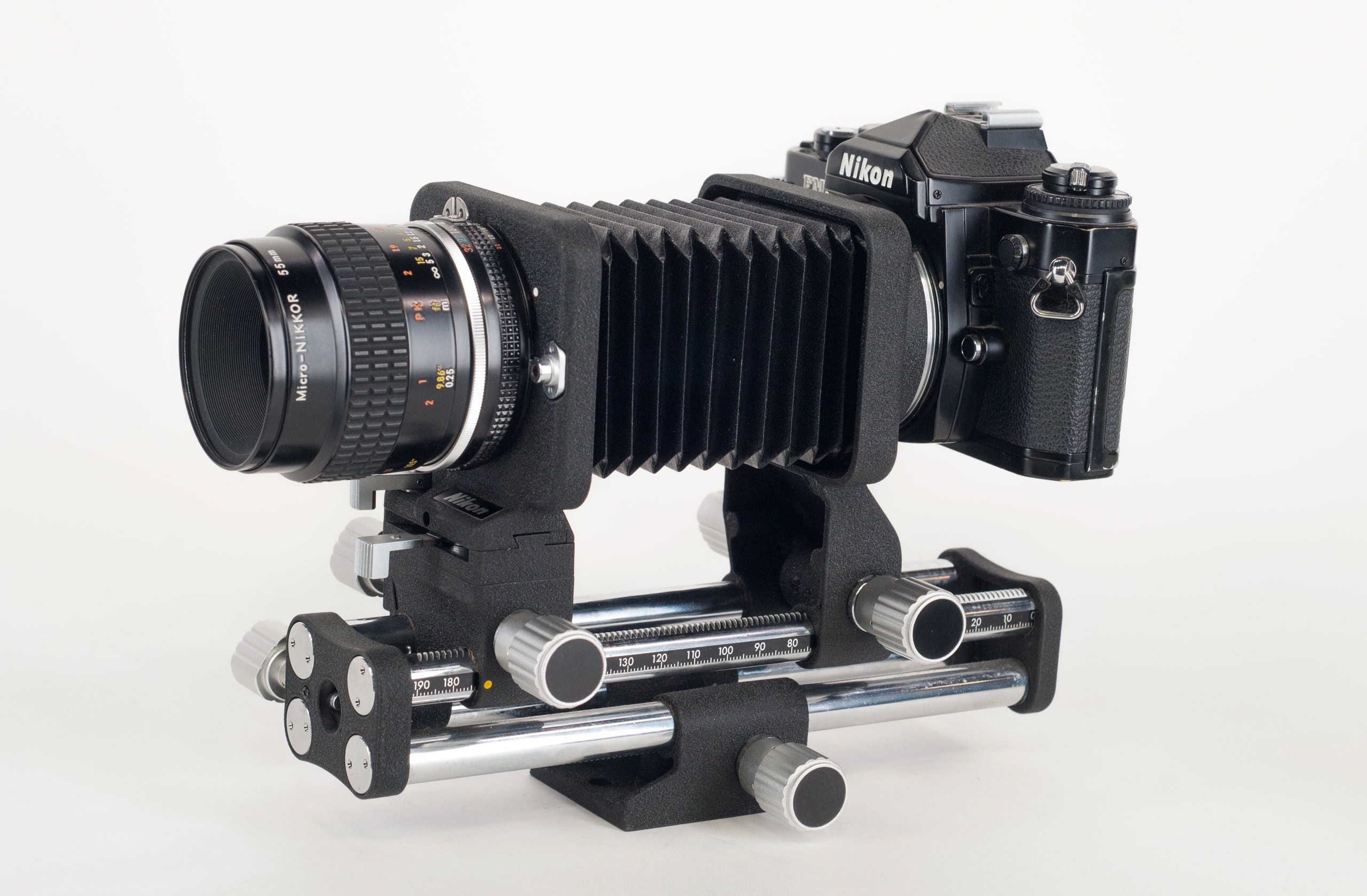 A digital camera with bellows, reversal rings, macro couplers, and focusing mechanism, typically used for extreme closeups of exceedingly small objects. Photo credit B&H Photo
 A digital camera’s closeup macro rings.
 A tube lens of the type used for extreme closeup photos of exceedingly small objects. Photo credit B&H Photo
The photographer used a digital camera “to which he attached a bellows, a tube lens and a microscope objective (basically a very high-powered magnifying glass).” The process requires that the “subject insect … be divided into as many as 20 or 30 sections, each of which is photographed separately.” Lighting is critically important “because eyes, legs, wings, etc. reflect light differently ….” The photographer consequently “devised different lighting regimes for each.”
Even more remarkably:
The macrophotography camera setup has an extremely shallow depth of field, so the lens is electronically advanced toward the subject in seven-micron increments (a micron equals one millionth of a meter) taking 400 or 500 digital pictures that are then merged to create an image in sharp focus from front to back.
The article adds that, “Paradoxically, smaller insects require more files than larger ones.” In the aggregate, “the section images are joined to make a file that may be as large as eight, nine or 10 gigabytes … The detail is stunning ….”
 A research-grade microscope.
A topically related story was posted by the journal Nature, March 10, 2023, titled, “Gigantic map of fly brain is a first for a complex animal.” First published in the journal Science, the project was made possible when:
Researchers spent a year and a half capturing images of the brain of a single six-hour-old Drosophila larva with a nanometre-resolution electron microscope. Using a computer-assisted programme, they then pinpointed the neurons and synapses and spent months manually checking them.
Science reported a related July 19, 2018, story titled, “In a ‘tour de force,’ researchers image an entire fly brain in minute detail.” The imaging methodology was, so to speak, mind-boggling:
[Researchers] soaked a fly’s brain in a solution containing heavy metals, which bind to the membranes of neurons and to proteins at the synapses …. [Next the team used] a diamond knife [to] cut the brain into about 7000 slices, each of which was struck with a beam of electrons from the microscope to create an image.
The process required a camera that could capture 100 frames per second, a robotic system to scoot each brain slice into place with nanometer precision, and software to stitch together the resulting 21 million pictures. The result is a reconstruction that lets researchers zoom in on the features of an individual synapse.
FROM THE SMALLEST TO THE LARGEST
The Orange County Register carried a similar article, February 18-19, 2023, titled, “See how the world’s largest photograph was created in Irvine [CA].” The picture involved 6 photographers working for 8 months to create a “panoramic view of … the former Marine Corps Air Station El Toro ….”
 A commercial, pinhole camera, sometimes used for stitching together individual photos to create a pantographic image of one very large object. Photo credit marekuliasz/Shutterstock
Using an ancient “pinhole camera” concept, the process captured “images of every single building” on the 4,800-acre base. The picture was created from about 500,000 individual images, shot from distances of 50-75 yards between the camera and the object being photographed. Each negative was then painstakingly exposed on one solid piece of muslin cloth, 31 feet tall and 111 feet long. The end-result was a single, giant, composite, historically significant photograph of virtually the entire military facility.
IMAGING IMPLICATIONS FOR NEUROLOGY IN HUMANS
The journal EHP, Environmental Health Perspectives, November 20, 2018, published an article titled, “The Brain before Birth: Using fMRI [functional magnetic resonance imaging] to Explore the Secrets of Fetal Neurodevelopment.” In this case, the imaging technology is used to study the trillions of neural connections, called the connectome, which link the “billions of threadlike fibers [that] crisscross the brain.”
The author explains that “fMRI is not perfect,” and that the images “generated by the technology must be manipulated to correct for distortion and to scale brain scans to a consistent, comparable template.” Equally problematic is the fact that “technical issues potentially result in artifacts that may not be recognized as errors.”
OCEANOGRAPHIC RELEVANCE
Arstechnica.com/science, May 17, 2023, posted an essay titled “3D ‘digital twin’ showcases wreck of Titanic in unprecedented detail.” Previous film of the wreck employed low-resolution cameras, and a 1997 documentary produced by James Cameron used “miniature models and special effects … since Cameron couldn’t get the high-quality footage he needed for a feature film.” The new pictures of the ship were obtained using two submersibles as camera platforms, mapping
… every millimeter of the wreck, including the debris field spanning some three miles. The result was a whopping 16 terabytes of data, along with over 715 still images and 4K video footage. That raw data was then processed to create the 3D digital twin. The resolution is so good, one can make out part of the serial number on one of the propellers.
‘This model is the first one based on a pure data cloud, that stitches all that imagery together with data points created by a digital scan, and with the help from a little artificial intelligence, we are seeing the first unbiased view of the wreck.’
One of the producers explained to BBC News that “you have to map every square centimeter—even uninteresting parts, like on the debris field you have to map mud, but you need this to fill in between all these interesting objects.”
 Submersible research craft with digital imaging equipment, generally used for deep sea exploration and composite photography. Photo credit maliao/Shutterstock
On the same topic, a May 17, 2023 insider.com story, headlined “First-ever full 3D scan of the Titanic on the sea bed reveals the ruined ocean liner in incredible detail,” describes the production as “the largest underwater scanning project in history.” It explains that “Previous footage has only allowed you to see one small area of the wreck at a time,” but this “model will allow people to zoom out and look at the entire thing for the first time ….”
METEOROLOGICALLY RELATED IMAGE PROCESSING
The Wall Street Journal, February 17, 2023, ran a story headlined “El Niño Likely to Form By Summer ….” It described the process by which weather forecasts are also made using composite imagery:
NOAA meteorologists make predictions of the ENSO cycle using statistical models that compare historical records with current ocean and atmospheric conditions. They also use computer models that combine data from satellites, ocean buoys, ships’ weather balloons and land-based stations into algorithms that form a picture, or map of what the future state of the atmosphere looks like based on the model’s calculations.
 A weather satellite of the type currently used to collect meteorological data.
IMAGE PROCESSING MAKES INVISIBLE DEEP SPACE PHENOMENA VISIBLE
According to gizmodo.com, on July 12, 2022, “the first full-color images from the Webb Space Telescope showed countless nebulae, galaxies, and a gassy exoplanet as they had never been seen before.” The article, headlined “Are the Colors in Webb Telescope Images ‘Fake’?” answers an emphatic “NO!” As it turns out, “Webb only collects infrared and near-infrared light, which the human eye cannot see ….”
The story goes on to explain that:
Image developers on the Webb team were tasked with turning the telescope’s infrared image data into some of the most vivid views of the cosmos we’ve ever had. They assign various infrared wavelengths to colors on the visible spectrum, familiar reds, blues, yellows, etc. But while the processed images from the Webb team aren’t literally what the telescope saw, they’re hardly inaccurate.
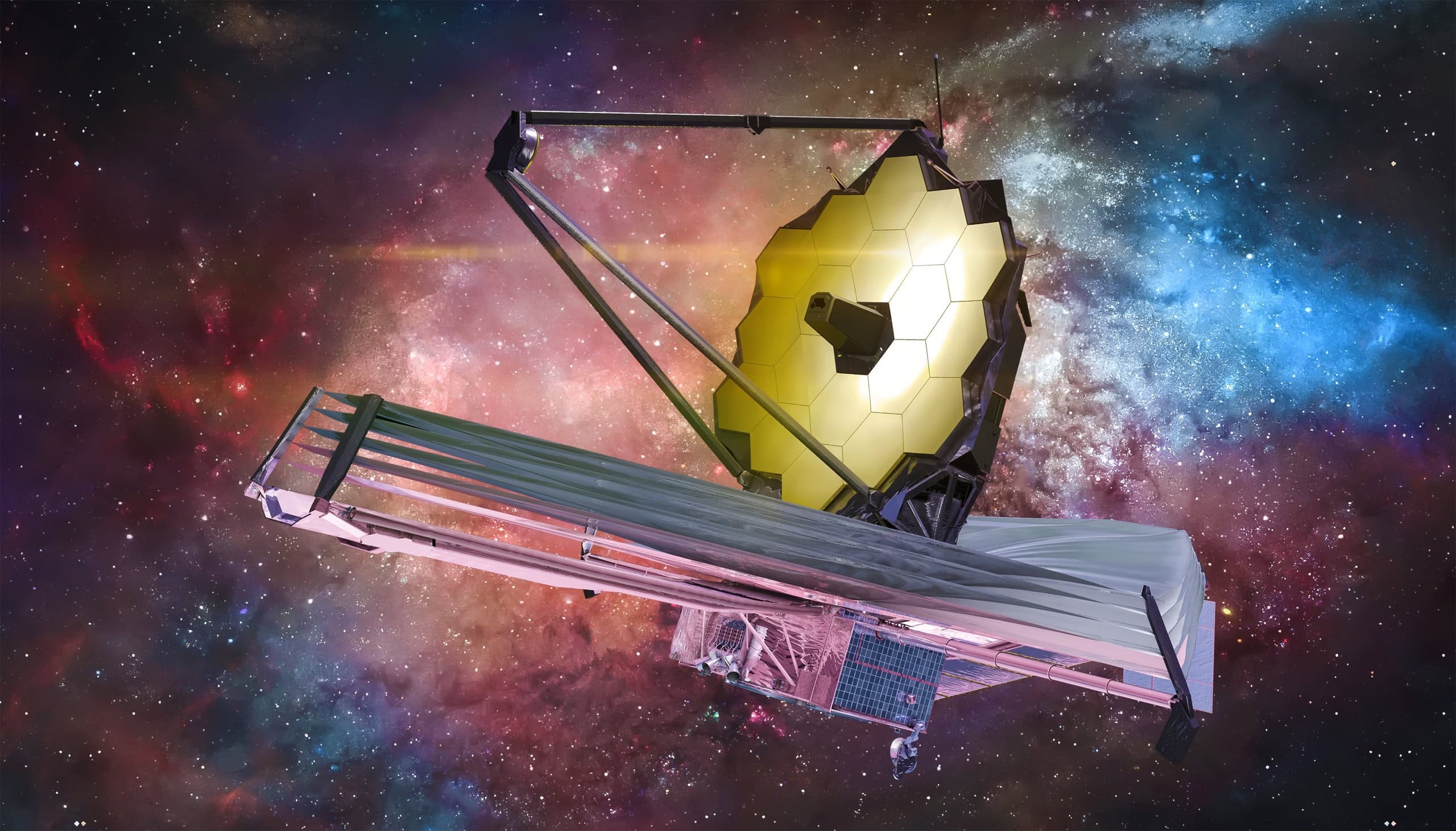 James Webb satellite telescope used for extensively processed, deep space imaging.
By way of further explanation, “Astronomy is often done outside the visible spectrum, because many of the most interesting objects in space are shining brightly in ultraviolet, x-rays, and even radio waves.” Instruments such as Webb were “designed to extend the power of our vision, to go beyond what our eyes are capable of doing, to see light that our eyes are not sensitive to.”
Using infrared light to image objects enables astronomers to “penetrate thick clouds of gas and dust in space, allowing researchers to see previously hidden secrets of the universe,” and then convert that monochromatic light into wavelengths visible to the human eye. Even more image processing is then necessary because “Webb’s raw images are so laden with data that they need to be scaled down before they can be translated into visible light.”
The Wall Street Journal, July 13, 2023, published a spectacular image of the “Rho Ophiuchi cloud complex, the closest star-forming region to earth … seen in a composite of separate exposures acquired by the James Webb Space Telescope using its NIRCam instrument.” As described above, the original image was monochromatic, but later processed to colorize it, and thereby make it visible to the human eye.
 An image of the center of the Milky Way galaxy
The New York Times, April 23, 2023, published a story on astronomical imaging which detailed the technology used to digitally enhance deep space photography. Headlined “Mysteries Residing at the Magnetic Heart of the Milky Way,” the article describes a “new celestial image” with “an impressionistic swirl of color” which “represents a first step toward understanding the role of magnetic fields in the cycle of stellar death and rebirth. The resulting map “reveals previously invisible details of a stretch of the central Milky Way 500 light-years wide” by assigning colors which “represent different temperatures of interstellar dust: Green indicates cool, dense dust; pink indicates warmer dust.”
The arbitrary assignment of color to reveal otherwise hidden details also plays a role in “showing the directions of magnetic force in the clouds.” Yellow streaks,” for instance, “are jets of hot ionized gas, which emits radio waves.” A specialized spectrograph “measured the direction of polarization of the infrared light emanating from the dust, revealing the directions of the magnetic fields point by point.” The story concludes by noting that “Every new generation of sees a new version of our galaxy,” because imaging technology continues to evolve to produce pictures far beyond anything discernable by the unaided human eye.
An even more arcane imaging technology is described in a Wall Street Journal article dated July 3, 2023, and titled “Launched Space Telescope To Study Dark Universe.” The story details the process by which “dark matter” is imaged:
Only 5% of the universe is made of matter that we can see [ironically, according to NOAA, the National Oceanic and Atmospheric Administration, only 5% of the earth’s oceans have been explored]. Now the European Space Agency’s Euclid mission will help probe the rest of what’s out there – what scientists call dark matter, which holds the cosmos together, and dark energy, which is responsible for our universe expanding …. Both are undetectable using traditional telescopes and astronomy.
 A satellite telescope of the type
The imaging challenge which must be overcome to create the intended “picture” is that dark matter “neither emits or absorbs light,” it can only be detected indirectly, “by observing the effects of its gravity in space.” That indirect process involves Euclid sensing “the position and shape of a distant galaxy, [and the fact that] the light from that cosmic structure will get bent ever so slightly by the dark matter between it and the telescope, and scientists can note that deviation.”
So whether we look up or down, in or out, many of the most important features of our universe can only be seen, at a cosmic or even sub-atomic level, by applying exceedingly complex image processing technologies.
Shareable Items
Stock Image Resources
Many stock image resources are available across the internet. These links provide quick access to embryonic and fetal imagery available for free or for purchase.
About ERF
The Education Resource Fund is a non-profit, 501(c)(3) science foundation, which facilitates the creation and distribution of a broad range of instructional materials authored and produced by individuals and organizations whose branded and copyrighted projects (domestic and international) advance the state of knowledge in subject areas vital to the public interest.
Contact ERF
The Education Resource Fund
PO Box 3950
Laguna Hills, CA 92654