
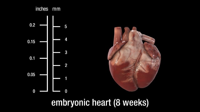
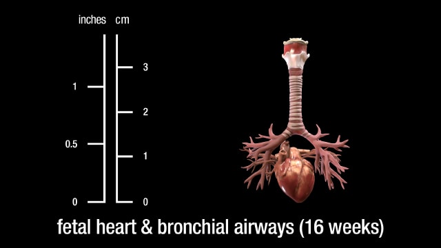
The heart is the first functional organ in vertebrate embryos.
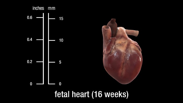
Heart development (also known as cardiogenesis) refers to the prenatal development of the heart. This begins with the formation of two endocardial tubes which merge to form the tubular heart, also called the primitive heart tube. The heart is the first functional organ in vertebrate embryos.
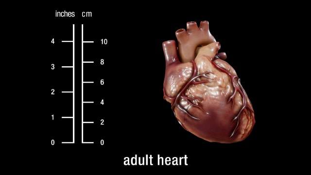
The heart is a muscular organ in most animals, which pumps blood through the blood vessels of the circulatory system. Blood provides the body with oxygen and nutrients, as well as assisting in the removal of metabolic wastes. In humans, the heart is located between the lungs, in the middle compartment of the chest.
During weeks 8 to 10, the cerebrum begins its development in earnest. Neurons proliferate and begin their migration throughout the brain. The anterior commissure, which is the first interhemispheric connection (a small one), also develops. Reflexes appear for the first time during this period.
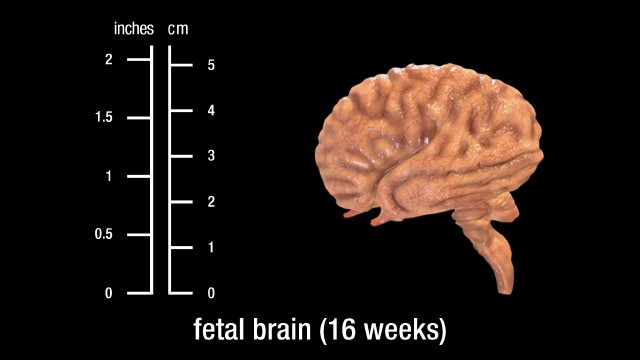
In the second trimester, the fetal brain begins to direct the compression of the chest muscles and movement of the diaphragm. These are like practice breaths and are controlled by the brainstem. Sucking and swallowing begin around week 16, and by week 21 the fetus can swallow amniotic fluid.
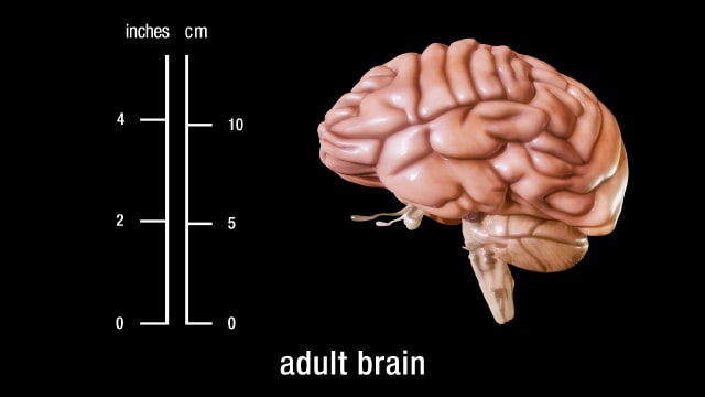
The brainstem provides the main motor and sensory nerve supply to the face and neck via the cranial nerves. Of the thirteen pairs of cranial nerves, ten pairs (or twelve, if the diencephalon is included in the brainstem) come from the brainstem. The brainstem is an extremely important part of the brain as the nerve connections of the motor and sensory systems from the main part of the brain to the rest of the body pass through the brainstem. This includes the corticospinal tract (motor), the dorsal column-medial lemniscus pathway (fine touch, vibration sensation, and proprioception), and the spinothalamic tract (pain, temperature, itch, and crude touch). The human brain is the central organ of the human nervous system, and with the spinal cord makes up the central nervous system.
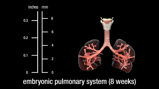
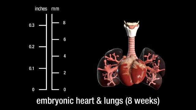
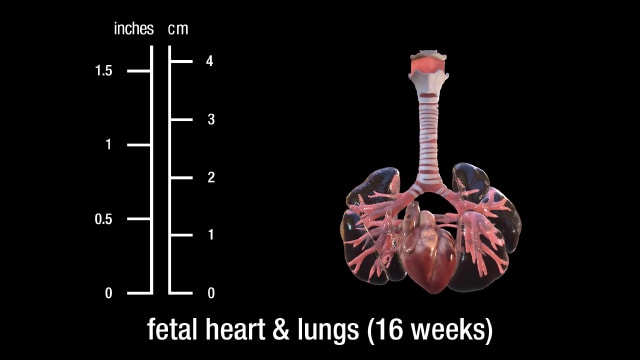
Stages of normal human lung development: During the first stage (0-7 weeks, embryonic stage), the lung arises as a ventral diverticulum of the primitive foregut endoderm with the lobar and segmental bronchi appearing at the fifth week and arteries and veins developing as avascular buds. There then follows the pseudoglandular stage (8-17 weeks) when branching of the airways and vessels takes place.
The respiratory system has conducting and gas exchange components, with the bifurcating airways and accompanying pulmonary arteries conducting air and blood to peripheral capillary-lined airspaces for gas exchange.
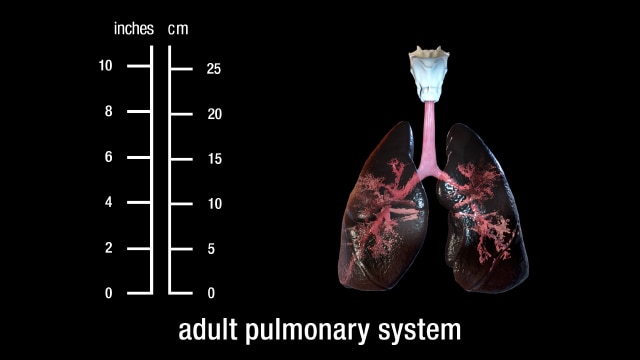
The pulmonary system includes the lungs, the bronchial tree, and the pulmonary vessels. The typical respiratory rate for a healthy adult at rest is 12–18 breaths per minute. The respiratory center sets the quiet respiratory rhythm at around two seconds for an inhalation and three seconds exhalation. This gives the lower of the average rate at 12 breaths per minute.
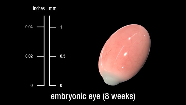
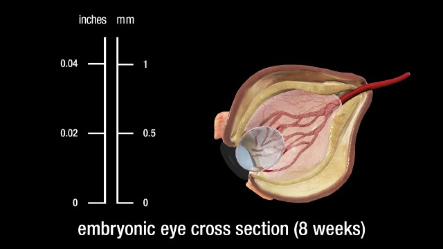
The eyes have moved forward on the face. At 8 weeks, the eyelids start to form and fuse together to protect the other developing eye structures.
Although eye development can be considered to commence around day 22, when the optic sulci (optic primordium) appear as shallow grooves or pits in the inner aspect of the neural plate or neural folds and the embryo is around 2 mm in length with eight somites, the group of cells that constitute the eye primordium or eye field have already begun to express a set of eye field transcription factors (EFTFs).
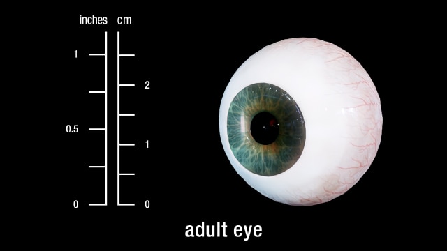
The human eye is a sensory organ, part of the sensory nervous system, that reacts to visible light and allows us to use visual information for various purposes including seeing things, keeping our balance, and maintaining circadian rhythm.
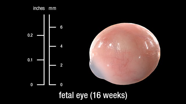
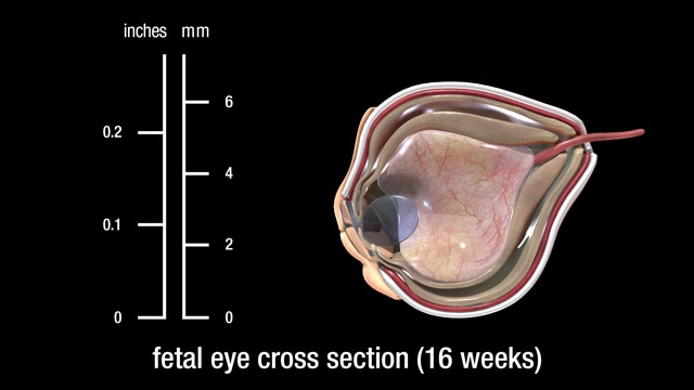
Cells differentiate into specific types according to their projected role in the visual process, such as rods and cones, with axons from the retinal ganglion cells forming the optic nerve. It takes six months for the retinal layers to grow from the neural ectoderm. The macula needs four or five months just to begin, and it will mature six months after birth.
Although eye development can be considered to commence around day 22, when the optic sulci (optic primordium) appear as shallow grooves or pits in the inner aspect of the neural plate or neural folds and the embryo is around 2 mm in length with eight somites, the group of cells that constitute the eye primordium or eye field have already begun to express a set of eye field transcription factors (EFTFs).
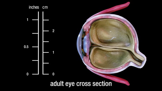
Eyes are organs of the visual system. They provide living organisms with vision, the ability to receive and process visual detail, as well as enabling several photo response functions that are independent of vision. Eyes detect light and convert it into electrochemical impulses in neurons. In higher organisms, the eye is a complex optical system which collects light from the surrounding environment, regulates its intensity through a diaphragm, focuses it through an adjustable assembly of lenses to form an image, converts this image into a set of electrical signals, and transmits these signals to the brain through complex neural pathways that connect the eye via the optic nerve to the visual cortex and other areas of the brain.
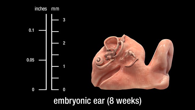
The ear is a composite structure with multiple embryonic origins , the external and middle ears arise from the first and second pharyngeal arches and the intervening pharyngeal cleft , membrane and pouch.
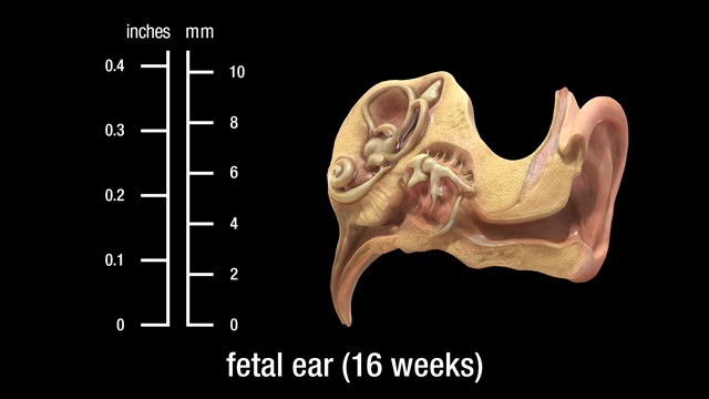
The ear is a composite structure with multiple embryonic origins. The external and middle ears arise from the first and second pharyngeal arches and the intervening pharyngeal cleft, membrane, and pouch.
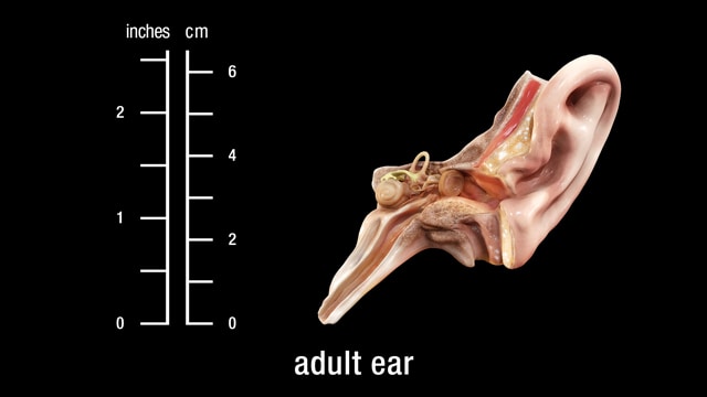
The ear is the organ that enables hearing and, in mammals, body balance using the vestibular system. In mammals, the ear is usually described as having three parts—the outer ear, the middle ear and the inner ear. The outer ear consists of the pinna and the ear canal. The middle ear includes the tympanic cavity and the three ossicles. The inner ear sits in the bony labyrinth, and contains structures which are key to several senses: the semicircular canals, which enable balance and eye tracking when moving; the utricle and saccule, which enable balance when stationary; and the cochlea, which enables hearing. The ears of vertebrates are placed somewhat symmetrically on either side of the head, an arrangement that aids sound localization.
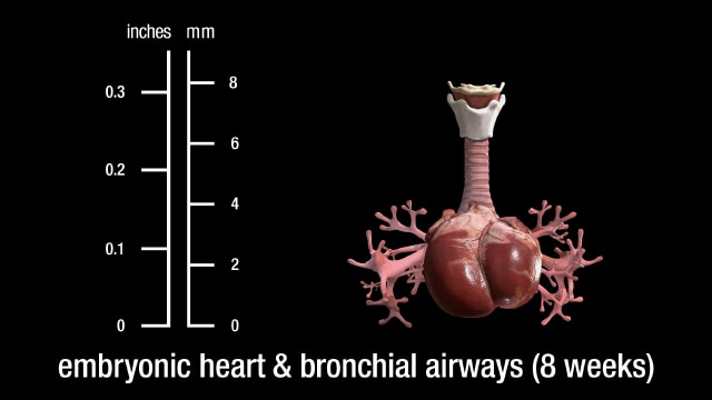
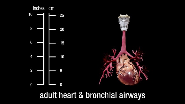
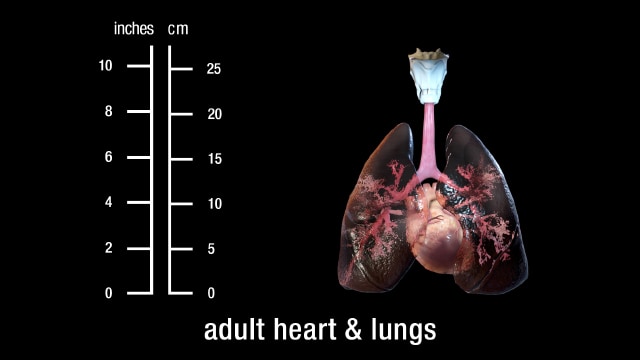
Heart development (also known as cardiogenesis) refers to the prenatal development of the heart. This begins with the formation of two endocardial tubes which merge to form the tubular heart, also called the primitive heart tube. The heart is the first functional organ in vertebrate embryos. The respiratory system has conducting and gas exchange components, with the bifurcating airways and accompanying pulmonary arteries conducting air and blood to peripheral capillary-lined airspaces for gas exchange.
Heart development (also known as cardiogenesis) refers to the prenatal development of the heart. This begins with the formation of two endocardial tubes which merge to form the tubular heart, also called the primitive heart tube. The heart is the first functional organ in vertebrate embryos. The respiratory system has conducting and gas exchange components, with the bifurcating airways and accompanying pulmonary arteries conducting air and blood to peripheral capillary-lined airspaces for gas exchange.
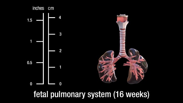
A bronchus is a passage or airway in the lower respiratory tract that conducts air into the lungs. The first or primary bronchi to branch from the trachea at the carina are the right main bronchus and the left main bronchus. These are the widest bronchi, and enter the right lung and the left lung at each hilum. The main bronchi branch into narrower secondary bronchi or lobar bronchi, and these branch into narrower tertiary bronchi or segmental bronchi. Further divisions of the segmental bronchi are known as 4th order, 5th order, and 6th order segmental bronchi, or grouped together as subsegmental bronchi. The bronchi, when too narrow to be supported by cartilage, are known as bronchioles. No gas exchange takes place in the bronchi.
Stages of normal human lung development: During the first stage (0-7 weeks, embryonic stage), the lung arises as a ventral diverticulum of the primitive foregut endoderm with the lobar and segmental bronchi appearing at the fifth week and arteries and veins developing as avascular buds. There then follows the pseudoglandular stage (8-17 weeks) when branching of the airways and vessels takes place.
Heart development (also known as cardiogenesis) refers to the prenatal development of the heart. This begins with the formation of two endocardial tubes which merge to form the tubular heart, also called the primitive heart tube. The heart is the first functional organ in vertebrate embryos. The respiratory system has conducting and gas exchange components, with the bifurcating airways and accompanying pulmonary arteries conducting air and blood to peripheral capillary-lined airspaces for gas exchange.
The normal resting adult heart rate is 60–100 beats per minute. The typical respiratory rate for a healthy adult at rest is 12–18 breaths per minute. The respiratory center sets the quiet respiratory rhythm at around two seconds for an inhalation and three seconds exhalation. This gives the lower of the average rate at 12 breaths per minute.
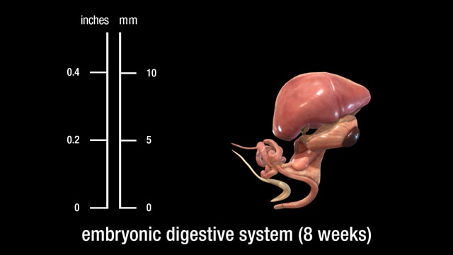
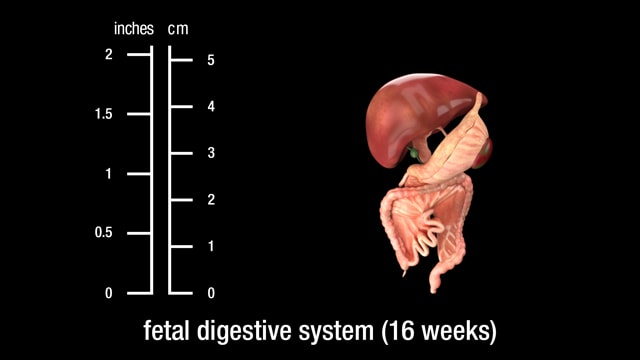
Both the epithelium and the parenchyma of glands associated with the digestive tract (e.g., liver and pancreas) are derived from endoderm. The muscular walls of the digestive tract (lamina propria, muscularis mucosae, submucosa, muscularis externa, adventitia and/or serosa) are derived from splanchnic mesoderm.
As a result of cephalocaudal and lateral folding of the embryo, a portion of the endoderm derived from gastrulation is incorporated into the embryo to form the primitive gut.
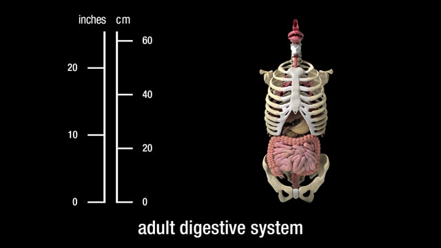
The digestive system plays a fundamental role ensuring liquids we ingest are broken down into useful nutrients and chemicals. The digestive tract consists of the mouth, or oral cavity, with its teeth, for grinding the food, and its tongue, which serves to knead food and mix it with saliva. Then, there is the throat, or pharynx; the esophagus; the stomach; the small intestine, consisting of the duodenum, the jejunum, and the ileum; and the large intestine, consisting of the cecum, a closed-end sac connecting with the ileum, the ascending colon, the transverse colon, the descending colon, and the sigmoid colon, which terminates in the rectum. Glands contributing digestive juices include the salivary glands, the gastric glands in the stomach lining, the pancreas, and the liver and its adjuncts – the gallbladder and bile ducts.
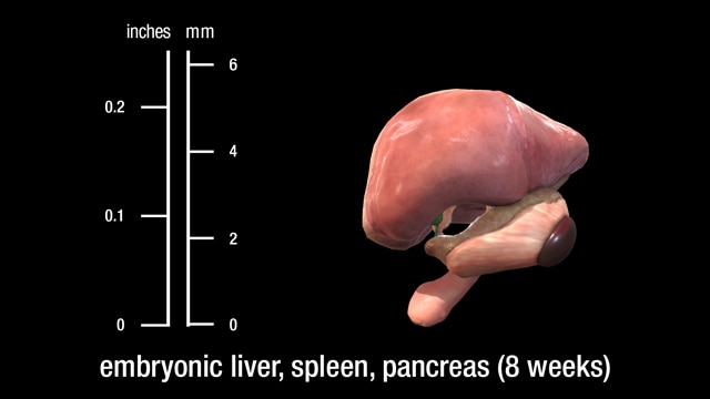
Both the epithelium and the parenchyma of glands associated with the digestive tract (e.g., liver and pancreas) are derived from endoderm. The muscular walls of the digestive tract (lamina propria, muscularis mucosae, submucosa, muscularis externa, adventitia and/or serosa) are derived from splanchnic mesoderm.
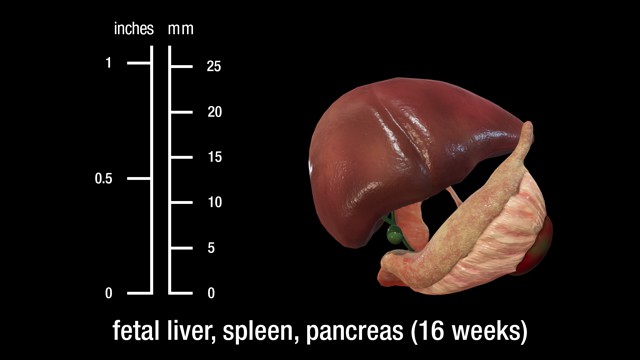
The liver, gallbladder, and biliary duct system arise as a ventral outgrowth from the caudal end of the foregut early in the fourth week of development. This outgrowth, known as the hepatic diverticulum, extends into the septum transversum, which is the future diaphragm. The hepatic diverticulum grows rapidly and divides into cranial and caudal portions. The larger cranial portion forms the primordium of the liver parenchyma. Proliferating endodermal cells develop into cords of hepatocytes and into the epithelial lining of the biliary system.
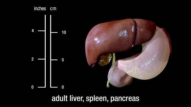
The pancreas and bile duct (biliary) systems together form an important part of the digestive system. The pancreas and liver produce juices (pancreatic juice and bile) which help in the process of digestion.